Download
1 / 57
581 likes | 925 Views
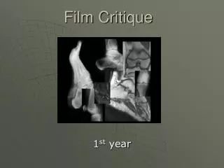
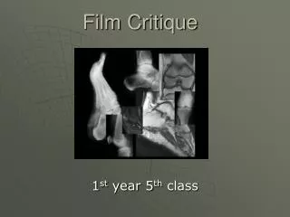
Film Critique. 1 st year 5 th class. Toes Standard views. *AP *Oblique (medioblique) *Lateral (mediolateral/lateromedial). Structures shown.

E N D
Film Critique 1st year 5th class
ToesStandard views *AP *Oblique (medioblique) *Lateral (mediolateral/lateromedial)
Structures shown • AP projection of the phalanges of the foot (*1st toe only has 2 phalanges the 2nd-5th have 3 phalanges) ***We need from distal phalanx to the distal end metatarsal.
AP Right 1st Toe Sesamoids
Check the film for: • No rotation of phalanges • Interphalangeal and metatarsophalangeal joint spaces open (no bent toes) • Toes should be separated from each other so there is no soft tissue overlap • Soft tissue and bony trabeculation (this is to check for a good technique)
AP left 1st toe Rotation of toe Soft tissue overlap AP
Structures shown **** We do a medioblique: • An oblique projection of the phalanges • The Interphalangeal joints and 2nd -5th metatarsophalangeal joints open ***Distal phalanx to the distal end of the metatarsal • Toes should be separated from each other • Both soft tissue and bony trabeculation should be seen (techn)
Oblique left 2nd toe LT Cadaver bone
Lateral left 1st toe • Might need tape, straw or tongue depressor to separate toes
Lateral toes Lateral left 3rd toe Lateral left 2nd toe lateromedial Mediolateral • Do lateromedial (1st-3rd) and mediolateral (3rd-5th) to get the toe closest to the film
Structures shown: Lateral toe • A lateral projection of the phalanges: Phalanges in profile (toenail should appear lateral) • The interphalangeal joints spaces open. The MTP joints will be overlapped but may be seen in some patients. ***The distal phalanx to the distal ends of the metatarsals • Phalanx, without superimposition of adjacent toes. When superimposition cannot be avoided, the proximal phalanx must be demonstrated. • Toes should be separated from each other • Soft tissue and bony trabeculation (techn)
Lateral left 2nd toe Lateral left 1st toe
Tongue depressor Lateral Left 2nd toe
Foot • Standard views *AP * AP Oblique (medioblique) *Lateral (mediolateral)
AP Right Foot Intermediate Base of the 5th Common area for a foot fracture base of 5th Jones fracture
AP Right foot **In this view you Will not see the Calcaneus!!
Structures shown: • Dorsoplanter (AP) projection of the tarsals anterior to the talus, the metatarsals,and the phalanges • You will not see the whole calcaneus on this view. Why? • Some people angle 10 degrees toward the heel on this view ***You want all of the phalanges, metatarsals and tarsals distal to the talus on your image
Check film for: • Motion • Rotation: there will be overlap of second- fifth metatarsal bases • Open joint space between medial and intermediate cuneiform • No overlap of toes • Density- are the toes burned out
Oblique Right Foot medioblique
Structures shown: • AP medioblique projection of the phalanges and metatarsals • Interspaces open between the cuboid and calcaneus, the cuboid and the 4th and 5th metatarsals, the cuboid and the lateral cuneiform and the talus and the navicular • Cuboid is in profile • Sinus tarsi, calcaneus, navicular,& base of the fifth are seen
Oblique Left Foot Calcaneus?
Check for: • Enough rotation when the 3rd – 5th metatarsals bases are free from superimposition • The lateral tarsals with less superimposition than in the AP • Joint spaces open • Base of the fifth metarsal is seen • Density: are the toes seen and are the tarsal seen • Tip of toes to calcaneus on the image
Lateral Right Foot R • mediolateral
Structures shown: • Mediolateral projection of the entire foot. ***You need distal ends of the tib/fib, ankle joint, calcaneus to the distal phalanges.
Check for: • Tip of toes to calcaneus and distal tib/fib on the image • Metatarsals nearly superimposed • Density to see toes, metatarsals and tarsals
Good Positioning Poor : heel not flat Poor : knee elevated Poor : foot not flat
CALCANEUS • Standard views *AP axial (plantodorsal) *Lateral (mediolateral)
Sustentaculum tali Trochlear process tuberosity
Structures shown: • An axial projection of the calcaneus ***from the tuberosity to the sustentaculum tali and trochlear process
Check for: • Calcaneus should be visualized to include the talocalcaneal joint • No rotation of calcaneus (check the first or fifth metatarsals) • Density to see joint without burn out of tuberosity (two films if not using DR or CR)
Rotation / foot flexion Rotation : can see 4th & 5th metatarsals Too much flexion Can’t see joint space Good
Structures shown: • Lateral projection of the ankle joint and the calcaneus and adjacent tarsals.
Check for: • No rotation of the calcaneus • Density can you see soft tissue and bone • Sinus tarsi seen • Ankle joint and adjacent tarsals should be on the film
Ankle • Standard views *AP *OBL (mortise) *Lateral (mediolateral)
Structures shown • AP projection of the ankle joint, ***distal ends of tib/fib and the proximal portion of the talus
Check for: • Talotibial joint space should be seen • Ankle joint should be centered • Moderate over lapping at the tibiofibular articulation is normal ***Area from the distal tibia and fibula to the talus should be included
Structures shown: • Distal ends of the tib/fib with the entire ankle mortise joint demonstrated in profile.(all three sides of the mortise joint should be open.)
AP OBLIQUE ANKLE Too much AP The entire ankle mortise joint should be demonstrated in profile. We oblique 15-20 degrees to open all three joints.