Introduction to Animal Diversity: Exploring the Kingdom
320 likes | 337 Views
Discover the vastness of the animal kingdom and their diverse characteristics. From multicellular organisms to specialized cells and body plans, learn about the evolutionary history and development of animals.

Introduction to Animal Diversity: Exploring the Kingdom
E N D
Presentation Transcript
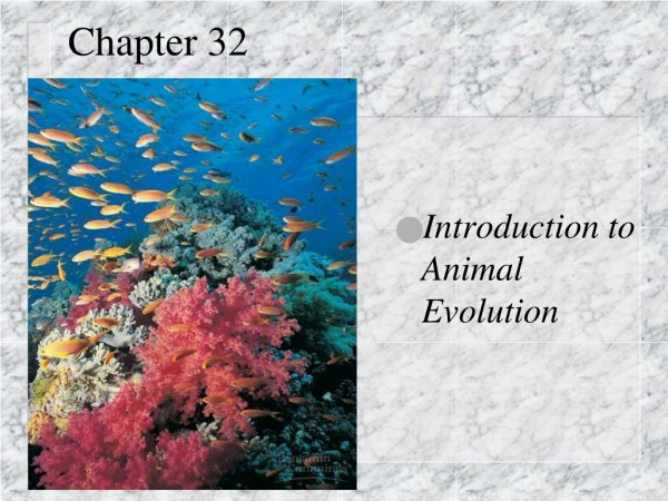
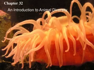
Chapter 32 • An Introduction to Animal Diversity
Figure 32.1 • Welcome to Your Kingdom • The animal kingdom extends far beyond humans and other animals we may encounter
Animal are multicellular, heterotrophic eukaryotes with tissues that develop from embryonic layers
Nutritional Mode • Heterotrophs ingest their food
Cell Structure and Specialization Multicellular eukaryotes, Cells lack cell walls
Bodies are held together by structural proteins, e.g. collagen • Nervous tissue and muscle tissue
Reproduction • Most animals reproduce sexually • Diploid stage dominating the life cycle
Development • Sperm fertilizes egg zygote cleavage blastula gastrulation formation of embryonic tissue layers gastrula
In most animals, cleavage results in the formation of a multicellular stage called a blastula. The blastula, a hollow ball of cells. Only one cleavage stage–the eight-cell embryo–is shown here. The zygote of an animal undergoes a succession of mitotic cell divisions called cleavage. 2 3 1 Blastocoel Cleavage Cleavage The endoderm of the archenteron de- velops into the tissue lining the animal’s digestive tract. 6 Cross section of blastula Eight-cell stage Blastula Zygote Blastocoel Endoderm The blind pouch formed by gastru- lation, called the archenteron, opens to the outside via the blastopore. 5 Ectoderm Gastrulation Gastrula Blastopore Most animals also undergo gastrulation, a rearrangement of the embryo in which one end of the embryo folds inward, expands, and eventually fills the blastocoel, producing layers of embryonic tissues: the ectoderm (outer layer) and the endoderm (inner layer). 4 • Early embryonic development in animals Figure 32.2
Hox genes regulate development of body form • Hox family of genes has been highly conserved, yet produces a wide diversity of animal morphology
The history of animals spans more than a billion years • Great diversity of living species and even greater diversity of extinct ones
Single cell Stalk • Common ancestor of living animals • Lived ~1 billion million years ago • May have resembled modern choanoflagellates, protists that are the closest living relatives of animals Figure 32.3
Digestive cavity Somatic cells Hollow sphere of unspecialized cells (shown in cross section) Reproductive cells Colonial protist, an aggregate of identical cells Beginning of cell specialization Infolding Gastrula-like “protoanimal” • Was probably a colonial, flagellated protist Figure 32.4
(b) (a) Neoproterozoic Era (1 Billion–524 Million Years Ago) • Early members of the animal fossil record Figure 32.5a, b
Paleozoic Era (542–251 Million Years Ago) • The Cambrian explosion • Earliest fossil appearance of many major groups of living animals • Several current hypotheses Figure 32.6
Mesozoic Era (251–65.5 Million Years Ago) • Dinosaurs were dominant terrestrial vertebrates • Coral reefs emerged, becoming important marine ecological niches for other organisms
Cenozoic Era (65.5 Million Years Ago to the Present) • Mass extinctions of terrestrial and marine animals • Modern mammal orders and insects diversified
(a) Radial symmetry. The parts of a radial animal, such as a sea anemone (phylum Cnidaria), radiate from the center. Any imaginary slice through the central axis divides the animal into mirror images. Body Plans • Radial symmetry • Like in a flower pot Figure 32.7a
(b) Bilateral symmetry. A bilateral animal, such as a lobster (phylum Arthropoda), has a left side and a right side. Only one imaginary cut divides the animal into mirror-image halves. • Bilateral symmetry • two-sided symmetry Figure 32.7b
Bilateral animals • Dorsal (top) side and a ventral (bottom) side • Right and left side • Anterior (head) and posterior (tail) ends • Cephalization, the development of a head
Tissues • Collections of specialized cells isolated from other tissues by membranous layers
Animal embryos • Form germ layers, embryonic tissues, including ectoderm, endoderm, and mesoderm • Diploblastic animals • two germ layers • Triploblastic animals • three germ layers
Body Cavities • In triploblastic animals • body cavity may be present or absent
(a) Coelomate. Coelomates such as annelids have a true coelom, a body cavity completely lined by tissue derived from mesoderm. Body covering (from ectoderm) Coelom Tissue layer lining coelom and suspending internal organs (from mesoderm) Digestive tract (from endoderm) Figure 32.8a Coelom • A true body cavity derived from mesoderm
Body covering (from ectoderm) (b) Pseudocoelomate. Pseudocoelomates such as nematodes have a body cavity only partially lined by tissue derived from mesoderm. Muscle layer (from mesoderm) Pseudocoelom Digestive tract (from ectoderm) Pseudocoelom • Body cavity not lined w/ mesoderm Figure 32.8b
Body covering (from ectoderm) Tissue- filled region (from mesoderm) Digestive tract (from endoderm) (c) Acoelomate. Acoelomates such as flatworms lack a body cavity between the digestive tract and outer body wall. Acoelomates • No body cavities Figure 32.8c
Protostome and Deuterostome Development • Distinction based on early development
Deuterostome development (examples: echinoderms, chordates) Protostome development (examples: molluscs, annelids, arthropods) (a) Cleavage. In general, protostomedevelopment begins with spiral, determinate cleavage.Deuterostome development is characterized by radial, indeterminate cleavage. Eight-cell stage Eight-cell stage Spiral and determinate Radial and indeterminate Cleavage • Protostomes • Cleavage is spiral and determinate • Deuterostomes • Cleavage is radial and indeterminate Figure 32.9a
(b) Coelom formation. Coelom formation begins in the gastrula stage. In protostome development, the coelom forms from splits in the mesoderm (schizocoelous development). In deuterostome development, the coelom forms from mesodermal outpocketings of the archenteron (enterocoelous development). Coelom Archenteron Coelom Mesoderm Blastopore Mesoderm Blastopore Enterocoelous: folds of archenteron form coelom Schizocoelous: solid masses of mesoderm split and form coelom Figure 32.9b Coelom Formation • Protostomes • Splitting of mesoderm coelom (schizocoelous development) • Deuterostomes • Enterocoelous development
Mouth Anus Digestive tube Anus Mouth Mouth develops from blastopore Anus develops from blastopore Figure 32.9c Fate of the Blastopore • Protostomes • Blastopore becomes the mouth • Deuterostomes • Blastopore becomes the anus
Rotifera Cnidaria Porifera Annelida Mollusca Chordata Phoronida Nemertea Ctenophora Nematoda Arthropoda Ectoprocta Brachiopoda Echinodermata Platyhelminthes “Radiata” Deuterostomia Protostomia Bilateria Eumetazoa Metazoa Ancestral colonial flagellate • Hypothesis of animal phylogeny based on anatomy Figure 32.10
Cnidaria Chordata Mollusca Annelida Rotifera Silicarea Phoronida Nemertea Calcarea Arthropoda Ctenophora Ectoprocta Brachiopoda Nematoda Echinodermata Platyhelminthes “Radiata” Deuterostomia Lophotrochozoa “Porifera” Ecdysozoa Bilateria Eumetazoa Metazoa Ancestral colonial flagellate • Hypothesis based on molecular data Figure 32.11