Download
1 / 12
120 likes | 230 Views
This study introduces a novel method for cardiac motion and partial volume correction in 3D PET imaging, leveraging a mass conservation-based optical flow approach. We analyze data from 14 patients with coronary heart disease (CHD) using gated and ungated PET acquisition techniques. The method significantly improves the precision of motion correction in cardiac phases by addressing common imaging challenges such as blur and noise. Our results demonstrate accurate correlation in myocardial thickness and blood pool activity, showcasing its potential for enhanced cardiac imaging diagnostics.

E N D
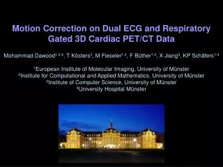
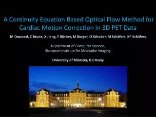
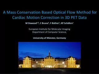
A Mass Conservation Based Optical Flow Method for Cardiac Motion Correction in 3D PET Data M Dawood1,2, C Brune2, F Büther1, KP Schäfers1 European Institute for Molecular Imaging Department of Computer Science, University of Münster, Germany
Cardiac Motion and Partial Volume Coronal slice through non-attenuated PET 1 h. p. i., 18FDG, CHD patient Cardiacmotion Ungated data Large blur, low noise (Problem in plaque imaging) Onephase Small blur, highnoise
Step 1: Gating ECG signal … Series ofimagesreconstructedfromcardiacgated PET acquisition
Mass conservation in cardiac data Systole Diastole Mass Conservation
Visual result Cardiacphases All gatesdeformedto Diastole
Visual result Onephase Small blur, highnoiseNoise 36 Ungateddata Large blur, lownoiseNoise 25 All phasesmotioncorrected Small blur, lownoiseNoise 22
Quantitative results on patient data Data: 14 patients with known CHD ca. 4 MBq/Kg body weight 18F-FDG Scan time ca. 15 minutes, 1:15 hours post injection Listmode acquisition on Siemens Biograph 16 scanner Quatification methods: Correlation of ROI (40x40x40) with target phase Myocardial thickness. FWHM of Gaussian fit to line profile Mean activity in blood pool in LV
Quantitative results 1: Correlation of end-systolic gate with target phase
Quantitative results 2: Myocardial thickness End-systole End-diastole End-systole after MC
To conclude: A method for cardiac motion and partial volume correction was presented. The results on patient data show that the motion was corrected precisely. Thankyou