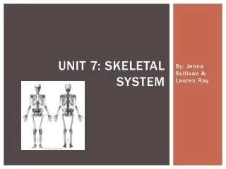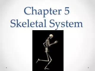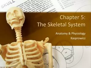
Understanding the Skeletal System: Structure, Function, and Ossification Processes
The skeletal system serves crucial functions including support for soft tissues, protection of delicate organs, and enabling movement. It comprises cartilage and bone, with cartilage providing a flexible framework while bone offers strength and structure. Bone types include compact and spongy bone, and their formation involves processes like intramembranous and endochondral ossification. The periosteum, a dense layer surrounding bones, aids in nutrition and repair. Different bone classifications exist based on shape, each tailored for specific functions within the human body.

Understanding the Skeletal System: Structure, Function, and Ossification Processes
E N D
Presentation Transcript
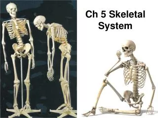
Skeletal system Composed of cartilage and bone
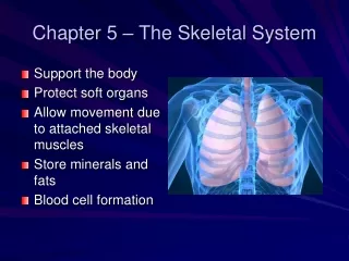
Functions • Support soft tissue • Prtects delicate organs • Movement • Storage area ( Calcium, phosphorus, fats • Hemopoiesis: blood cell formation
Cartilage: • consists of chondrocytes living in a cavity (lacuna) and is avascular with cells being nourished by diffusion
Bone • consist of osteocytes living in lacunae and a great deal of non-living matrix with collagen fibers • However is highly vascularized • The matrix contains 33% collagenous fibers and 67% mineral salts ( calcium phosphate, calcium carbonate called hydroxy-apatites) • Embryonic skeleton is all cartilage but slowly ossifys
Bone structures • Diaphysis: the shaft ( compact bone) • Epiphysis: ends ( thin layer of compact bone covering spongy bone • Metaphysis: region where epiphysis and diaphysis come together, region of epiphyseal plate
Bone structures • Articular cartilage: thin hyaline cartilage covering of epiphyseal surface at joints • Medullary cavity (marrow cavity): space within diaphysis
Bone structures • Periosteum: dense irregular, white fibrous layered covering of a bone. • Is composed of 2 layers: • Fibrous layers: outer layer composed of bibrous connective tissue containing blood vessels • Osteogenic layer: layer closer to bone that contains elastic fibers, blood vessels, osteoclast and osteroblast
Functions of periosteum • Growth • Repair • Nutrition • Attachments for ligament and tendons
Classification of bones by shape • Long: longer than they are wide ex: most extremities, phalanges • Short: wider than they are long ex: carpals and tarsals • Flat: ex: cranium, ribs, shoulder girdle • Irregular: vertebrae and facial • Sessamoid: patella • Wormian: grows between bones of skull
2 Types of bone tissue • Compact: • Covers spongy bone, is dense and provides strength • Structural unit of compact bone is the osteon
Osteon • The osteon is a hard bone matrix arranged in rings (lamellae) around a haversian canal • Volkman’s canal: carry blood vessels and nerves from the periosteum into the haversian canal and on into the medullary cavity • Osteocytes: mature osteoblasts, live in the lacunae between lamellae
Canaliculi: are tiny canals which connect lacunae with each other and with the haversian canal. • Inside canaliculi are cellular extensions of osteocytes creating a network for nutrients and distribution • Areas between osteons where the lamellae are only partially circled are called interstitial lamellae. • They are the remnants of old partially destroyed osteons.
Types of bone tissue • Spongy bone • Consists of a lattice work of bone called trabeculae • In between the trabeculae are spaces filled with red bone marrow and blood vessels • There is no network of canals because the blood can reach the osteocytes by diffusion through marrow spaces • Is less dense and less strong and is found in the epiphysis of long bones and the bulk of flat bones or irregular bones
Bone formation ( osteogenesis or ossification • Embryo’s skeleton is made of cartilage and fibrous membranes • At around 8 weeks after conception and throughout childhood ( until about age 20) this connective tissue model is slowly ossified ( turned to bone)
Intramembraneous ossification • Results in formation of flatbones and some irregular ones by ossification of pre existing fibrous connective tissue • The osteoblasts begin secreting bone matrix eventually forming spongy bone …later the outer layers are reconstructed into compact bone
Intramembraneous ossification • The original connective tissue which encloses the “growing” bone becomes the periosteum • Some osteoblasts trap themselves in the matrix (lacunae) and are now called osteocytes • Bones are destroyed and reformed many times before adulthood and final size and shape
Endochondral ossification • Most bones formed this way • Involves replacement of hyaline cartilage with bone • Embryonic skeletons are cartilage enclosed by connective tissue called perichondrium • Once a blood vessel penetrates the perichondrium of the embryonic bones the chondrocytes become osteoblasts which begin forming bone matrix
Endochondral ossification • Perichondrium is now periostium • Area where bone begins to form is called primary ossification center (central located on bone) • Between ages 1-5 blood vessels enter epiphyses and secondary ossification centers occur (at birth ends of bones are still cartilage) • So primary and secondary ossification centers grow toward each other, replacing cartilage with bone.
Endochondral ossification • The cartilage between these ossification centers is called epiphyseal plate (don’t ossify until about 18-25) • Growth in diameter is due to osteoblasts in periosteum adding new osseous tissue to outside surface of bones • Initially all bone growth begins with spongy bone which is later remodeled into compact bone.
Bone Remodeling • The continuous balance between bone formation and bone reabsorption, occurs at all periosteal and endosteal surfaces • Bone is constantly replacing itself throughout adult life.
Bone disorders • Rickets: childhood disease due to lack of vitamin D (sunlight) causes cartilage not to be calcified, bones are soft and malformed • Osteomalacia: adult rickets… too little vitamin D • Solution: both need dietary calcium and sunlight (vitamin D) • Both disorders can also be due to either kidney or liver disease and resultant is a poor processing of calcium
Bone disorders • Osteoporosis: character sized by a decrease in bone mass due to decease osteoblast activity. Generally age related ( decrease in hormone activity, females often due to menopause) • Also due to insufficient exercise, resulting in brittle porous bones • Bones are easly broken, decreased height, hunched back etc… • Solution: exercise, calcium and vitamin supplements and maybe hormones
Bone disorders • Osteomyelitis: includes all infectious disease of bone
Divisions of skeletal system • Axial skeleton: forms midline of skeleton • 80 of 206 bones in human body are axial ( 28 in skull alone • Appendicular: remiainng 126 bones • Found in arms, legs, pectoral and plevic girdle
Fractures: breaks in bones • Simple: Bone breaks cleanly, but does not penetrate skin • Compound; broken ends of bone protrude trough skin • Comminuted: bone fragments into many pieces • Compression: bone is crushed
Depression: broken bone portion is pressed inward • Impacted: broken bone ends are forced into each other • Spiral: ragged break occurs when excessive twisting forces are applied to a bone • Greenstick: bone breaks incompletely, much in the way a green twig breaks
Colle’s fracture: fracture of distal end of lateral forearm (radius) in which distal fragment is displaced posterior • Stress fracture: microscopic fissures in bone that form without evidence of injury to other tissue no visible break. • Pott’s fracture: fracture at distal end of lateral legbone (fibula)) with serious injury of distal tibial articulation
4 steps of fracture repair • Hematoma: swelling, blood clotting, influx of fibroblasts and granulation tissue • Fibrocartilagineous callus: collagen fibers bridge broken gap forming a callus • Bony callus: collagen is calcified • Remodeling: of spongy to compact bone and removal of excess callus
Articulations (joints) • Functional classification • synarthroses: immovable joint (skull sutures) • Amphiarthroses: slightly moveable (between vertebrae or in pelvis) • Diarthrosis: freely moveable
Structural Classification • Fibrous joints: no join cavity, bones held together by fiberous connective tissue • Cartilaginous joints: still no cavity, bones held together by cartilage • Synovial joints: have cavities, free moving • Have articular cartilage (haline) • Have articular capsule (units articular bones) • Has synovial fluid • Has reinforcing ligaments
Types of Fibrous joints • Sutures: only in skull, bones touch joined by connective tissue (synathrotic) • Syndesmosis: bones slightly separated (amphiarthrotic) ex tibia, fibula • Gomphosis: peg in a socket ex teeth