Download
1 / 62
650 likes | 728 Views
Learn about mandible fractures, their evaluation, and treatment options. Understand the epidemiology and management techniques for favorable outcomes.

E N D
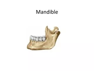
Mandible Fractures Jacques Peltier MD Matthew Ryan MD UTMB – Dept of Otolaryngology May 2004
History • Edwin Smith Papyrus 1650 described Hx, Phy, Diagnosis. Often fatal disease • Hippocrates – Described monomaxillary dental fixation and binding • Sulicetti – 1492 Described “tie teeth of jaw to teeth of uninjured jaw”
History • Schede 1888 – Bone plate of steel secured with 4 screws • Luhr 1960 – Developed mandibular compression plates • Michelet and Champy 1970’s – Placement of small bendable non-compression plates
Epidemiology • Mandible most common after nasal fractures • Mandible : Zygoma : Maxilla 6:2:1 • Ellis 4711 facial fractures, 45% with mandible fractures • Assault>MVA>Fall>Sports
Epidemiology • Sites of weakness • Third molar (esp. impacted) • Socket of canine tooth • Condylar neck
Epidemiology • Boole et al (laryngoscope) 5196 fractures • Young military men • Angle 35%, Symphysis 20%, Body 12%, Condylar 9%, Subcondylar 4%, Ramus 4%, Alveolar 3%, Coronoid 1% • 70% 1 fracture, 30% 2 fractures, .2% more than 2 • Facial lacs 30%, other facial fx. 16%, C-spine 0.8%
Favorable vs. Unfavorable • Masseter, Medial and Lateral Pterygoid, and Temporalis tend to draw fractures medial and superior • Almost all fractures of angle unfavorable
Evaluation • Stabilization via ATLS protocol • Part of secondary survey • Pain, malocclusion, trismus, V3 sensory deficit • History of TMJ (earlier mobilization) • Blow to face favors parasymphyseal fracture and contralateral angle fracture • Fall to chin (bilateral condylar fractures)
Evaluation • Previous occlusion (Class I-III) • Psychiatric, nutritional, gastrointestinal, seizure disorders • Previous facial trauma • Other injuries (c-spine, intra-abdominal, likely prolonged intubation)
Physical Exam • Complete Head and Neck exam • Palpable step off • Tenderness to palpation • Malocclusion • Trismus (35 mm or less) • FOM hematoma • Altered sensation of V3 • Crepitus
Physical Exam • Dental Exam • Lost, fractured, or unstable teeth • Dental Health • Relation to fracture • Quantity
Physical Exam • Unilateral fractures of Condyle • Decreased translational movement, functional height of condyle • Deviation of chin away from fracture, open bite opposite side of fracture Bilateral fractures of condyle - Anterior open bite
Evaluation • Panorex, mandible series • CT scan • Not as diagnostic as plain films for nondisplaced fractures of mandible. • Most useful for coronoid and condylar fractures, associated midface fractures
Physiology • Primary Healing • In rigid fixation techniques • Lag screws, compression plates, Recon plate, external fixation, Wire fixation, Miniplate fixation • No callus formation • Question of bone resorption
Physiology • Secondary bone healing • Callus formation • Remodeling and strengthening • MMF, Wire fixation, Miniplate fixation
Closed Reduction • Favorable, non-displaced fractures • Grossly comminuted fractures when adequate stabilization unlikely • Severely atrophic edentulous mandible • Children with developing dentition
Closed Reduction • Length of MMF • De Amaratuga – 75% of children under 15 healed by 2 weeks, 75% young adults 4 wks • Juniper and Awty – 82% had healed at 4 wks • Longer period for edentulous fractures 6-10wks
Closed Reduction • Edentulous fractures • Bradley found absent inferior alveolar artery in 40% 60-80 yo’s • Periosteal blood supply disturbed by stripping • Up to 20% non-union despite type of treatment • May consider Gunning Splints
Open Reduction • Displaced unfavorable fractures • Mandible fractures with associated midface fractures • When MMF contraindicated or not possible • Patient comfort • Facilitate return to work
Open Reduction • Contraindications • General Anesthetic risk too high • Severe comminution and stabilization not possible • No soft tissue to cover fracture site • Bone at fracture site diffusely infected (controversial)
Open Reduction • Associated condylar fracture • Associated Midface fractures • Psychiatric illness • GI disorders involving severe N/V • Severe malnutrition • To avoid tracheostomy in patients who need postoperative intubation
Open Reduction • Intraosseous wiring • Semirigid fixation • Cheap • Technically difficult • Primary and Secondary bone healing
Open Reduction • Lag Screws • Rigid fixation (Compression) • Good for anterior mandible fractures, Oblique body fractures, mandible angle fractures • Cheap • Technically difficult • Injury to inferior alveolar neurovascular bundle
Open reduction • Ellis 41 patients with anterior lag screw technique • 4.9% infection rate • No malocclusion • No Non-union
Rigid Fixation • Compression plates • Rigid fixation • Allow primary bone healing • Difficult to bend • Operator dependent • No need for MMF
Rigid Fixation • Miniplates • Semi-rigid fixation • Allows primary and secondary bone healing • Easily bendable • More forgiving • Short period MMF Recommended
Rigid Fixation • Schierle et al studied experimental model, then applied in patients. • Model suggested two plates more stable • Patients divided into two groups with equal complication rates, equal functional results
Rigid Fixation • Reconstruction Plates • Good for comminuted fractures • Bulky, palpable • Difficult to bend • Locking plates more forgiving
External Fixation • Alternative form of rigid fixation • Grossly comminuted fractures, contaminated fractures, non-union • Often used when all else fails
Edentulous Fractures • Chalmers and Lyons 1976 – Recommended closed reduction to preserve periosteal blood supply • Chalmers and Lyons 1995 • 167 fractures in edentulous mandibles • ORIF 82% • 15% complications • 12% Fibrous union
Edentulous Fractures • ORIF • Inferior alveolar canal more superior in location • Vertical height 20mm compatible with standard plating systems • Vertical height 10mm or less, likely need rib graft • Plate removal after fracture healing if interferes with denture placement