Download
1 / 17
190 likes | 684 Views
Muscles of mandible. By: Abdul Muhaimin (D11 A007) Reference: Veterinary anatomy of domestic animal (Book). Mandibular muscles. Comprise muscle of mastication and superficial muscles of mandibular space. Innervated by mandibular nerve (trigeminal nerve)

E N D
Muscles of mandible By: Abdul Muhaimin (D11 A007) Reference: Veterinary anatomy of domestic animal (Book)
Mandibular muscles Comprise muscle of mastication and superficial muscles of mandibular space. Innervated by mandibular nerve (trigeminal nerve) Responsible for movement jaw (mastication) Covers mandibular space and hyoid apparatus ventrally. Different species have their own structure of muscle for mastication.
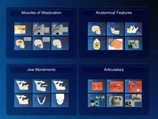
Mandibular muscle • Muscle of mastication • Masseter muscle • Medial and lateral pterygoid muscles • Temporal muscle • Superficial muscles of mandibular space • Digastric muscle • Mylohyoid muscle
Muscle of mastication • Masseter muscle • Broad multipennate muscle • Multiple tendinous intersections • Origin • From ventral border of zygomatic arch and facial crest • Insertion • On the lateral aspect of mandible • Extend • From facial notch to temporomandibular joint.
Masseter muscle • In carnivores, it is separated into 3 layers • Superficial, middle, deep (by tendinous sheets) • Superficial portion is the strongest • Middle layer is the weakest • In pig, these three layers are firmly fused • Tendinous intersections are pronounced, forming 5 distinct parts in the ox. • Change in fibre direction between each portion increases masticatory force of this muscle
Masseter muscle • In horse, it shows up to 15 tendinousintermuscular strands which are orientated sagitally and divide muscle into multiple layers. • Both sides masseter muscles act together will force upper and lower jaw together • Mandible can be move to the side of contracting muscle (grinding in herbivores)
Pterygoid muscle • Pass from palatine, pterygoid and sphenoid bones to the medial aspect of mandible. • Lateral pterygoid muscle is smaller than medial one. • In carnivores, both are fused at their origin • Horse: medial pterygoid muscle is covered by lateral one. Mandibular nerve passes across lateral surface of medial pterygoid muscle (separating 2 pterygoid muscles).
Pterygoid muscle • Pterygoid and masseter muscles contract bilaterally will raise mandible, but if working unilaterally, will draw mandible to side of contracting muscle. • Lateral portion also able to move mandible rostrally (mouth open)
Temporal muscle • Size varying in different species depending size fossa • The strongest muscle of head in carnivores. • Dolichocephalic dogs: temporal muscle meets corresponding muscle of opposite side in midline (form mid-line sulcus) • Brachycephalic dogs: two muscles don’t meet, no sulcus visible
Temporal muscle • Ruminant: temporal muscle is indistinct but visible in horse • Horse: temporal muscle however do not well developed compared to other masticatory muscles. • It raises mandible acting together with other masticatory muscles.
Superficial muscle of mandibular space • Assist muscles of mastication. Cover ventral side of lingual muscles in mandibular space. • Digastric muscle • Single bellied muscle in domestic animal (except horse) • Horse: has caudal and rostral bellies • Other domestic mammals: evolutionary bipartite structure is indicated by a fibrous intersection.
Digastric muscle • Rostral part is innervated by mylohyoid nerve (branch of mandibular nerve) • Caudal part by digastric branch of facial nerve • In carnivores: it is strong single-bellied muscle with delicate tendinous strands (mark division between rostral and caudal portion) • Ruminants: tendinous intersection between two bellies is indistinct. • Horse: rostral belly depresses mandible and opens mouth
Mylohyoid muscle • Form sling between inner surface of body of mandible • Innervated by mylohyoid nerve (branch of mandibular nerve), assigned to mandibular group • Can also be seen as lingual muscle (its function) • Supports tongue and raises it towards palate