Download
1 / 64
740 likes | 1.73k Views
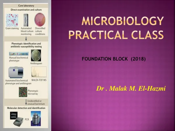
MICROBIOLOGY Practical Class. FOUNDATION BLOCK (2018). Dr . Malak M. El-Hazmi. MICROBIOLOGY. Laboratory diagnosis of infectious diseases. Microscopic examination. culture. Serological tests ( Ab ). Detection of Ag. Molecular method. Types of specimens. Bacteriology. Gram Stain.

E N D
MICROBIOLOGYPractical Class FOUNDATION BLOCK (2018) Dr . Malak M. El-Hazmi
Laboratory diagnosis of infectious diseases • Microscopic examination. • culture. • Serological tests (Ab). • Detection of Ag. • Molecular method .
Gram Stain G- bacilli
Gram negative cocci (Diplococci ) e.gNeisseria • Gram negative bacilli • e.g E. coli • Salmonella
Gram positive cocci in chain Streptococci Gram positive cocci in clusters Staphylococci Rx cloxacillin Cephalosporin if MRSA vancomycin Penicillin Cephalosporin
A gram-stained smear of a CSF sample from a 3 year old child seen in the emergency department presenting with fever and neck stiffness. Gram-positive diplococci & pus cells Streptococcus pneumoniae
This is a bacterium isolated from a child with sore throat and tonsillitis . A: Describe the Gram stain Gram positive B: Describe the shape and arrangement of the bacteria Cocci in chains
Following is the Gram stained smear of an organism isolated from a wound infection. Describe what you see in the slide above. Gram-positive cocci in clusters What is the likely organism ? Staphylococcus aureus
Following is the Gram-stained smear of from urethra of a 25 –year old male complaining of urethral discharge Describe the Gram stain of the intracellular bacteria Gram negative Describe the shape of the bacteria cocci ( diplococci)
Describe the Gram stain of this organism: Gram negative Describe its shape bacilli ( rods )
Bacteria culturing 1-Inoculation 3-Incubation 2-streaking Laboratory Incubator
Identification of streptococci by hemolytic reaction Beta-hemolytic Streptococcus colonies Alpha-hemolytic Streptococcus colonies Gamma-hemolytic Streptococcus colonies
Biochemical testings • To confirm • the identity • of bacteria. Antibiotic susceptibility testings
Automated instrument for identification and susceptibility testings
Blood agar This is a general culture medium used for culture of bacteria.
This culture was grown from a sputum specimen of a 60 year old man complaining of cough, fever and chest pain. α hemolytic streptococci on blood agar
Herpes simplex virus -1 : Herpesviridae Enveloped virus Icosahedral capsid d.s DNA genome Loose envelope
These are electron micrographs of a virus Herpes virus Q1: Name this virus Enveloped virus , Icosahedral capsid, d.s DNA genome Q2: Describe its structure.
Adenovirus : Adenoviridae Nonenveloped virus Icosahedral capsid d.s DNA genome Only V with fiber
This is an electron micrograph of a virus Adenovirus Q1: Name this virus • Nonenveloped virus, with fiber • Icosahedral capsid & d.s DNA genome Q2: Describe its structure.
Rabies virus: Rhabdoviridae Enveloped virus Helical capsid s.s RNA genome Bullet shape
This is an electron micrograph of a virus Q1: Name this virus • Rabies virus Enveloped virus , Helical capsid & s.s RNA genome Q2: Describe its structure.
Influenza Viruses : Orthomyxoviridae Enveloped V & spikes Helical capsid Segmented s.s RNA Pleomorphic shape
This is an electron micrograph of a virus Q1: Name this virus Influenza Viruses Enveloped Virus with spikes , Helical capsid ,Segmented s.s RNA Q2: Describe its structure
Ascarislumbricoides (roundworm) Ascaris adult