Download
1 / 79
800 likes | 1.01k Views
Explore detailed diagrams and descriptions of facial and neck regions, dental embryology, and histology. Learn about key structures such as zygomatic arch, oral cavity, mental foramen, and more.

E N D
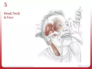
Face and Neck RegionsChapter 1 Dental Embryology, Histology, and Anatomy
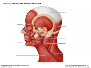
Regions of the face (Fig. 1-1) A C B D
Regions of the face (Fig. 1-1) A Reg. A Region B Region C C B Region D D
Regions of the face (Fig. 1-1) A. Zygomatic C. Oral B. Buccal D. Mental
Regions of the face (Fig. 1-2) E. Sternocleidomastoid muscle
Regions of the Face (Figure 1-2) F Zygomatic arch A G Parotid salivary gland
Frontal view of face (Fig. 1-3) H. I. J.
Frontal view of face (Fig. 1-3) H. I. J.
Frontal view of face (Fig. 1-3) H. Zygomatic arch I. Temporomandibular joint J. Buccal region
Nose (Fig. 1-4) L M K
Nose (Fig. 1-4) L Nasal septum M Ala K Nares (nostrils)
Buccal Region (Fig. 1-5) NQ NR NN NS NT NO _duct NP
Buccal Region (Fig. 1-5) Q R S N O T P
Frontal view of lips (Fig. 1-6) V W X U Y
Frontal view of lips (Fig. 1-6) V Philtrum W Vermilion X commissure U Tubercle Y zone Y border
Mandible (Fig. 1-8) Z 1 2 3
Mandible (Fig. 1-8) Z. Coronoid process 1.Condyle 2. Mental Foramen 3. Ramus
Neck region (Fig. 1-11) 5. 4. 6.
Neck region (Fig. 1-11) 5. Hyoid 4. Sternocleidomastoid 6. Thyroid
Lips Darby and Walsh, Dental Hygiene Theory and Practice 2nd Edition, Fig 12-6.
Oral Cavity and PharynxChapter 2 Dental Embryology, Histology, and Anatomy
Divisions of the Oral Cavity (Fig. 2-1) upper C D A towards cheek towards tongue E B lower F towards lip
Divisions of the Oral Cavity (Fig. 2-1) C Maxillary D Palatal A Buccal E Lingual B Facial / labial F Mandibular
Vestibule (Fig. 2-2) J G K H L M I mucosa
Vestibule (Fig. 2-2) J vestibule G Parotid papilla K Alveolar mucosa H Buccal mucosa L Muccobuccal fold M Vestibule I Labial mucosa
Describe appearance. • Smokeless Tobacco Keratosis Newland, Meiller, Wynn, and Crossley; Oral Soft Tissue Diseases, Lexi-Comp, Inc., 2001, p. 27
Vestibule (entranceway) Oral cavity proper (inside the teeth) Surfaces: Facial /labial Buccal Palatal Lingual Divisions of the Oral Cavity
Oral Vestibules N. Labial frenum • Oral mucosa (membrane) • Labial mucosa • Buccal mucosa • Alveolar mucosa • Labial frenum N. Labial frenum
Fordyce Granules (spots) • Ectopic sebaceous glands found in the buccal mucosa, labial mucosa, or vermillion zone • Multiple, small, white to yellow nodules Newland, Meiller, Wynn, and Crossley; Oral Soft Tissue Diseases, Lexi-Comp, Inc., 2001, p. 17
Clinical considerations wit h Oral Mucosa (Fordyce Granules with Linea Alba) Daniel and Harfst, Mosby’s Dental Hygiene, Concepts, Cases and Competencies, 2008, p. 298. Sebaceous fat glands
Abnormal condition of the buccal mucosa: Linea Alba (Frictional Keratosis)
Alveolar processes and permanent teeth Maxillary Mandibular
Tooth tissues (Fig. 2-5) What does the periodontal ligament (PDL) attach?
Alveolar Processes (Bones) (Fig. 2-6) • Alveolus (tooth socket) • Portions of gingiva
Mandibular Tori Darby and Walsh, Dental Hygiene Theory and Practice 2nd Edition, Fig 12-27.
Gingiva and Landmarks (Fig. 2-9) O Alveolar mucosa P Mucogingival juncion Q Attached gingiva
Gingiva and landmarks (Fig. 2-10) R S T (inside)
Gingiva and landmarks (Fig. 2-10) R Marginal gingiva S Interdentalgingiva/papilla T Sulcus
Oral cavity proper and boundaries (Fig. 2-11) Z U 1 V 2 3 4 5 W X 6 Y
Oral cavity proper and boundaries (Fig. 2-11) Z Fauces U Hard palate 1 Max. tuberosity V Soft palate 2 Pterygomandibular fold 3 Posterior faucial pillar 4 Palatine tonsil 5 Anterior faucial pillar W Uvula X Post. wall of pharynx 6 Retromolar pad Y Dorsal surface of tongue