Download
1 / 1
10 likes | 142 Views
Biotechnology Methods for Detecting Mercury Shaun Cote, Rebekah Pilling, Caryn K. Prudent é. Preliminary Data

E N D
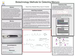
Biotechnology Methods for Detecting Mercury Shaun Cote, Rebekah Pilling, Caryn K. Prudenté Preliminary Data In the first phase of this research project, a mercury-protein bioconjugate was developed. An organomercury hapten was connected to a carrier protein, chicken immunoglobulin, via a linker arm. Mice were then immunized with this immunogen, causing the immune system to develop antibodies. Four mice were screened for antibody development, and the mouse with the highest titer was used to create mAbs. We were unsuccessful in developing the mAb, so testing will continue with the remaining three mice. [2]. Abstract Due to the high levels of mercury contamination in the environment and a need for better testing methods, we are exploring the synthesis of a mercury-protein bioconjugate to facilitate the development of a mercury antibody. Mice have been immunized with this mercury-protein bioconjugate and the antibodies will be collected for screening through Enzyme-Linked Immuno-Sorbent Assay (ELISA) to determine the effectiveness and sensitivity of the antibody. This poster will discuss the synthetic processes and structural analysis of our mercury-protein bioconjugate as well as the testing method by which we utilize the mercury antibody to detect the presence of mercury contamination Solid Phase Synthesis To develop a sensitive and selective ELISA to test with a monoclonal antibody (mAb), a solid phase must be developed to coat the polystyrene plates. To achieve maximum selectivity, a solid phase protein-mercury bioconjugate structurally different than the immunogen must be synthesized. I ran the synthesis through common organic pathways. First I reacted allyl amine with Di-tert-butyl dicarbonate to protect the amine. I then reacted the protected allyl amine with a mercury salt to attach mercury and an amine or alcohol to the allylic region of the amine. In my first reaction, I used mercury acetate (Hg(oAc)2) with an excess of allyl amine. This mercury salt gave a small yield, and the H1-NMR spectral data was compromised, as the resulting product had the same π bond as the starting material. In my second attempt at this reaction, I used a more reactive mercury salt, mercury nitrate (Hg(NO3)2), and 1,2 Pentane diol. The diol did not have a π bond, so H1-NMR was not compromised. To deprotect my t-Boc allyl amine structure, I used trifluoro acetic acid. This mercury group will be connected via a linker arm to a carrier protein, thus creating a solid phase structurally different than the immunogen Mercury Mercury is naturally found in environmental systems, but can be released into the environment through burning coal and other manufacturing processes. In the environment, mercury can combine with carbon to form methyl mercury, which is ingested through organisms, and works its way up through the food chain to humans. Mercury levels in environmental systems in Maine are among the highest in North America [1]. Synthesis Scheme Sandwich ELISA Assay + + Hg Hg Non-specific mercury antibody is washed away Mouse anti-mercury antibody trapped on solid phase Sample containing antibodies Immobilized solid phase (can vary) Enzyme + An antibody sandwich! Hg Enzyme Acknowledgments USM Faculty Senate Professors Ah-Kau Ng and Stephen Pelsue USM Chemistry Department References [1] Maine DEP; http://www.state.me.us/dep/mercury [2] Research done by Rebekah Pilling Enzyme labeled goat anti-mouse antibody Hg Adapted with permission from Caryn Prudente Polystyrene ELISA plates are coated with a solid phase containing the antigen of interest coupled to a protein or sugar. The inherent “stickiness” of the solid phase protein adheres it to the plate. A serum sample from immunized mice is then added to the ELISA plate. Mouse anti-mercury antibodies from the sample are retained on the plate as they bind to the mercury immobilized in the solid phase. Any non-mercury-specific antibodies are rinsed away. In a second step an enzyme labeled anti-mouse antibody is added to the plate and binds to the mouse anti-mercury antibodies. The enzyme labeled antibody can be used for detection and quantification. Once the ELISA is optimized if will provide a tool to detect and quantify mercury in various samples