
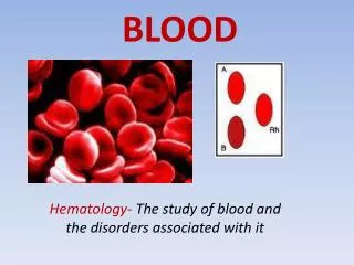
BLOOD
BLOOD. Hematology- The study of blood and the disorders associated with it. Blood Function. Blood is connective tissue that has 3 functions: 1. Transportation - oxygen, carbon dioxide, nutrients, heat, and hormones

BLOOD
E N D
Presentation Transcript
BLOOD Hematology- The study of blood and the disorders associated with it
Blood Function • Blood is connective tissue that has 3 functions: 1. Transportation- oxygen, carbon dioxide, nutrients, heat, and hormones 2. Regulation- The heat absorbing and coolant properties of water in blood plasma helps adjust body temperature 3. Protection- Blood clots to protect against excessive blood loss. It also carries white blood cells to fight infection.
Components of Whole Blood • 2 parts of blood: • Plasma- Liquid ≈55% of whole blood • Cells and cell fragments≈45% of whole blood * Hematopoeisis- The formation of blood cells and cell fragments * Hematocrit- The percentage of total blood that is occupied by red blood cells.
Plasma • Straw colored • 91.5% water, 7% proteins and 1.5% solutes • Solutes include nutrients, gases, enzymes, hormones, vitamins, and waste products • Plasma proteins include: • Albumins- help with the exchange of fluids across capillaries • Globulins- antibodies which fight infection • Fibrinogens-formation of blood clots
Red Blood Cells • Red blood cells- called erythrocytes • Biconcave disks • At maturity, do not have a nucleus nor organelles; therefore, cannot divide nor metabolize • Contain the protein hemoglobin. Each hemoglobin carries 4 iron molecules and each iron binds with 1 oxygen. Hemoglobin also gives blood its red color.
Red Blood Cells continued… • Lifespan is 120 days • Erythropoeisis- Formation of new red blood cells. Near the end of the process, red cells eject their nucleus, which causes the indentation • Anemia- lower than normal RBCs or hemoglobin • Hypoxia- Deficiency of oxygen • Signals for erythropoiesis to occur • The hormone that regulates erythropoiesis is erythropoietin which is produced by the kidneys. • Some athletes take erythropoietin to improve their performance. What are the dangers of this?
Blood Types • Blood typing is determined by the presence or absences of proteins on the surface of red blood cells. These proteins are called antigens. • Type A- has antigen A • Type B- has antigen B • Type AB- has both antigen A and B • Type O- does not have the A nor B antigen
Blood Types- draw these B A AB O
Antibodies • Antibodies are plasma proteins that bind to specific antigens to inhibit or destroy them. • People do not produce antibodies for the antigen on their red blood cell surface. If they did, the antibodies would attach to the antigens and cause the blood to clump. This clumping is called agglutination. • Antibodies bind with antigens because they fit together like a “lock and key”.
Blood Compatibility • When receiving blood from a donor, it is essential that the blood is compatible with the blood of the recipient. Otherwise, agglutination will occur, which could be fatal. • Compatibility is due to the antibodies that are present in the plasma of the recipient. • COMPLETE THE CHART
Rh Antigen • The Rh antigen is a separate antigen from the A & B antigen of red blood cells. It is called this because it was first discovered in the blood of the rhesus monkey. • If a person’s red cells have the Rh antigen, then they are typed “positive”. • If a person’s red cells lack the Rh antigen, then they are typed “negative”.
Blood Typing • SAFETY FIRST! • NO EATING NOR DRINKING!! • Wear gloves if you are going to puncture someone else’s finger. • Once you have used a lancet, fold it in half and put it in the red “Biohazard” bag. • Put all other possible contaminated items (used gloves, alcohol pads, and combi slide guides) in the “Biohazard” bag. • Clean off your area with a Clorox wipe when finished.
Blood Typing Procedure • Obtain the following items: combi slide guide, alcohol pad, disposable sterile lancet, red mixing spatula, blue mixing spatula, and yellow mixing spatula. • Vigorously swing the hand you will prick several times. • Wipe the middle finger with the alcohol pad and let it air dry. • Using the lancet, puncture the tip of the finger once. Immediately discard the lancet. • Squeeze your finger and wipe away the first drop of blood with an alcohol pad. • Force out additional blood by milking and put 1 drop on each of the 3 areas of the combi slide guide.
Blood Typing Procedure cont… 7. Place 1 drop of Anti-A, 1 drop of Anti-B and 1 drop of Anti-Rh next to the blood drops in the appropriate places. • Mix the blood and Anti solutions with the appropriate colored spatula. DO NOT CROSS CONTAMINATE! IMMEDIATELY DISCARD SPATULAS. • After 1 minute, look for clumping in your blood/Anti A&B mixtures. If clumping is present, then you have that antigen. • It is difficult to observe the results in the blood/Anti Rh mixture. Tilt the combi slide so that the Rh mixture runs to the side of the guide. Then look at the thin later of Rh mixture in the circle and observe your results.
Do our results agree with the national statistics? O+ (38.4%)A+ (32.3%)B+ (9.4%) O- (7.7%) A- (6.5%)AB+ (3.2%)B- (1.7%)AB- (0.7%)
From Blood Typing Worksheet • If an A+ person mates with a B- person, what are the possible genotypes of the children? You will need to do separate Punnett squares for the A/B antigens and the Rh antigen.
Another Genotyping Question • Is it possible for an O- mother and AB+ father to have a child who is B+? If yes, show the Punnett square that would support it.
Rh- Mothers • Rh- individuals do not produce Rh antibodies unless they are exposed to the Rh antigen. • If a Rh- mother gets pregnant with a Rh+ baby, there is no concern during pregnancy because red blood cells do not cross the placenta. • During delivery, however, baby blood might enter the mother’s system. Consequently, the mother will produce Rh antibodies. • This will create a problem if the mother becomes pregnant again later with a Rh+ baby. Her Rh antibodies can pass the placenta and cause agglutination in the baby. • To prevent this, Rh- mothers are always given a shot of antibodies at birth that will destroy any baby cells that enter her system and prevent her from producing her own antibodies.
White Blood Cells • Also called leukocytes • Have a nucleus and do not contain hemoglobin • Different types include neutrophils, eosinophils, basophils, lymphocytes and monocytes • The function of WBCs is to either destroy bacteria by phagocytosis (engulfing and digesting), combat the effects of allergic responses, create chemicals to intensify allergic reactions, or produce antibodies to inactivate antigens on viruses
More WBC facts.. • WBCs have antigens on the surface called major histocompatibility antigens (MHC). In addition to RBC antigens, MHCs are used to type tissue to identify compatible donors and recipients. • Less numerous than RBCs (outnumbered 700 to 1) • Life span ranges from days to years • Leukopenia is an abnormally low number of white blood cells (below 5000 cells/µl of blood)
Platelets • Cell fragments that do not contain a nucleus • Have a life span of 5-9 days
Hemostasis • Hemostasis is a sequence of responses that stops bleeding when blood vessels are injured. • Platelets form a plug at the injury site by sticking to parts of the damaged blood vessel. This is called platelet adhesion. • Platelets release chemicals that makes other platelets in the area sticky to create a large platelet mass. • Fibrin is a protein formed from plasma proteins that acts as a net to trap blood cells and create a clot at the injury site. This is called coagulation.
Hemophilia • A rare inherited disorder in which a person does not produce clotting proteins. Therefore, they have spontaneous nosebleeds, blood in the urine, hemorrhages in joints and tissue damage. • Hemophiliac patients are treated with transfusions of fresh plasma that contains clotting factors to relieve the tendency to bleed. • Video
Hematocrit Hematopoiesis Albumins Fibrinogens Hemoglobin Leukocytes Erythropoiesis Anemia Hypoxia Rh Major Histocompatibility Antigens Leukopenia 13. Agglutination 14. Type O 15. Coagulation 16. Erythropoietin 17. Erythrocytes 18. Globulins 19. Platelets 20. Hemostasis 21. Antibodies 22. Hemophilia 23. Plasma 24. Type AB “BLOOD”(instead of BINGO)


