Download
1 / 17
170 likes | 189 Views
Explore the structural dynamics of cellular components like microtubules, stress fibers, and intermediate filaments. Learn about centrosomes, polymerization regulation, muscle contraction, and more. Discover the intricate architecture vital for cell function.

E N D
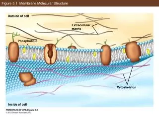
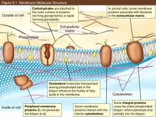
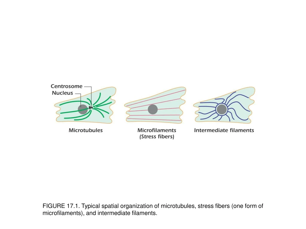
FIGURE 17.1. Typical spatial organization of microtubules, stress fibers (one form of microfilaments), and intermediate filaments.
FIGURE 17.2. The microtubule organizing center or centrosome consists of amorphous material enclosing a pair of centrioles.
FIGURE 17.3. Microfilaments and microtubules in fibroblasts grown in culture. The green shows microtubules radiating out from the microtubule organizing center while the red shows actin. (A) In this flattened cell the actin is organized as stress fibers. (B) In this rounded cell the actin is organized as a loose meshwork under the plasma membrane. The blue color is Hoechst staining and shows the nucleus. Ima-ges by Professor David Becker, University College London; used with permission.
FIGURE 17.6. Effects of taxol and colchicine on microtubules.
FIGURE 17.11. Actin polymerization is regulated by actin-binding proteins.
FIGURE 17.12. Integrins anchor the actin cytoskeleton to the extracellular matrix.
FIGURE 17.14. Amoeboid movement resembles the progression of a military tank.
FIGURE 17.15. Intermediate filaments are formed from rod-shaped monomers.
FIGURE 17.16. Anchoring junctions attach the cytoskeletons of adjacent cells.