Download
1 / 21
210 likes | 461 Views
HYDROSONOGRAPHY FOR THE DIAGNOSIS OF RESIDUAL TROPHOBLASTIC TISSUE. YARON ZALEL, MD. Sheba Medical Center, Israel. Incidence – 1%. Risk Factors – Termination of pregnancy (most common) Vaginal delivery

E N D
HYDROSONOGRAPHY FOR THE DIAGNOSIS OF RESIDUAL TROPHOBLASTIC TISSUE YARON ZALEL, MD Sheba Medical Center, Israel
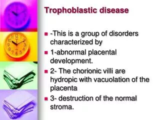
Incidence – 1% Risk Factors – Termination of pregnancy (most common) Vaginal delivery Caesarian Section Diagnosis – Diagnostic hysteroscopy - Only 70% detection rate
Hydrosonography - Intrauterine saline instillation May improve our ability to evaluate the intrauterine pathology
THE AIM To evaluate the accuracy, efficacy and safety of hydrosonography in diagnosing residual trophoblastic tissue
Material & Methods 1998 - 2000 23 patients – Vaginal bleeding and/or pelvic pains All patients treated with oxytocic agent Methergin (Methylergometrine maleate)
Material & Methods Hydrosonography – 8 French Foley catheter 3 ml balloon Plastic guide Catheter pulled back - ensure fixation - prevent spillage 2-5 ml saline Transvaginal sonography - Presence of lesion following instillation of saline Intralesional blood flow
Material & Methods Operative Hysteroscopy - Transcervical extraction of residual throphoblast Standard 26Fr, continuous Bow Urologic Resectoscope (with 24Fr cutting loop) Dilatation of cervix- Hegar 9 Distention & irrigation – 1.5% glycin solution Controlled by hysteromat
Results Pregnancy Types MA- 4 cases TOP - 4 cases CS - 1 case NVD - 3 cases
Results Hydrosonography – Echogenic intra- uterine lesion Group 2 Group 1 11 patient 12 patient Detached from the wall Adherent to uterine wall Blood flow- 0/11 Blood flow- 4/12
Hydrosonography & Residual throphoblast Post partum Post abortion
Results Histology Confirmed throphoblast tissue in 12 (100%) Gr 1 - Blood clots / decidua in 4 of Gr 2 (cont. bleeding / doctors’ concern)
Residual trophoblast Hydrosonography used for diagnosis- Endometrial polyps Sub-mucous myomas Thickened endometrium Tamoxyfen Infertility - endometrial cavity
Residual trophoblast Diagnosis of residua - Diagnostic hysteroscopy- only 70% accuracy (blood clots, decidua) Hydrosonography- - intra-uterine lesion not detached from uterine wall - 100% accuracy
Residual trophoblast Hydrosonography & Blood flow 4/12 in Gr. 1 Vs 0/11 in Gr. 2 P = 0.093 Presence - could add to diagnosis Absence - does not exclude (dead trophoblast)
Hydrosonography & Blood flow Post abortion Blood flow within lesion
Hydrosonography & Blood flow Post NVD
Residual trophoblast Advantages of hydrosonography Post partum - cervix opened - Inflated balloon lower risk of uterine rupture Uterine anomalies - location of lesion with regard to anomaly Mininal invasive, Easy to learn
Residual trophoblast Operative Hysteroscopy - Should follow hydrosonography Directed minimal curettage Little electricity Dilatation & Curettage - Blind non-directed Prevention of infection, scarring & Asherman’s
Hydrosonography - Conclusion Intrauterine lesion, not detached following saline instillation! Blood flow ? Easy to learn, Office procedure Especially useful post-partum High accuracy Operative hysteroscopy follows