Download

1 / 47
1.05k likes | 2.97k Views
Gestational Trophoblastic Disease. Yi-Chun Lee, MD Division of Gynecologic Oncology Department of Obstetrics & Gynecology State University of New York Downstate Medical Center. Introduction. A continuum of tumors arising in the fetal chorion during pregnancy Unique diseases

E N D
Gestational Trophoblastic Disease Yi-Chun Lee, MD Division of Gynecologic Oncology Department of Obstetrics & Gynecology State University of New York Downstate Medical Center
Introduction • A continuum of tumors arising in the fetal chorion during pregnancy • Unique diseases • the elaboration of beta-hCG • the sensitivity to chemotherapy • the immunobiological relationship between the disease and its host
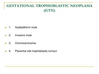
Introduction (cont.) • Histologically, GTD is classified as: • Hydatidiform mole (complete & partial mole) • Invasive mole (chorioadenoma destruens) • Choriocarcinoma • Placental site trophoblastic tumor • Clinically, GTD is classified as: • Benign trophoblastic neoplasia • Malignant trophoblastic neoplasia • non-metastatic • metastatic • good prognosis (low risk) • poor prognosis (high risk)
Hydatidiform Mole • Incidence • 7 to 10 times higher frequency of molar pregnancy in Asian countries than in North America or Europe • In the US: 1 in 1,500 live births • Ireland study from 1st & 2nd trimester abortions: • Incidence of complete mole: 1 : 1,945 • Incidence of partial mole: 1 : 695 • in Mexico: 1 in 200 pregnancies • in Taiwan: 1 in 125 pregnancies • ? due to reporting differences rather than true incidence differences • hospital-based vs. population-based data
Hydatidiform Mole • Risk factors • Socioeconomic & nutritional factors: • Low dietary carotene and animal fat • Vitamin A deficiency • Advanced maternal age: 5 to 10 folds greater risk when older than 40 • Not associated with partial mole • History of prior SAB and infertility • Relative risk of 3.1 for complete mole & 1.9 for partial mole among women with 2 or more prior miscarriages comparing to women with no SAB • Infertility: odds ratio of 2.4 for complete & 3.2 for partial mole • Repeat hydatidiform moles occur in 1.2 to 1.4% of patients • a subsequent greater risk of invasive mole or choriocarcinoma • No significant association with gravidity
Hydatidiform Mole • Etiology • Remains unclear • Appears to be due to abnormal gametogenesis and fertilization • The karyotype in 87% of hydatidiform mole is 46, XX • Most appear to arise from an anuclear empty ovum fertilized by a haploid sperm (23X), which then duplicates its own chromosomes • Up to 13% have 46, XY chromosomal composition • The result of two spermatozoa (23X+23Y) fertilizing an anuclear empty ovum • Whereas chromosomes in the complete mole are entirely of paternal origin, the mitochondrial DNA is of maternal origin
Diagnosis of Hydatidiform Mole • Primarily diagnosed in the 2nd trimester during the 1960s and 1970s, currently mostly diagnosed in the first trimester • Histologically • proliferation of the trophoblasts (both cytotrophoblast and syncytiotrophoblast) with varying degrees of hyperplasia and dysplasia • the chorionic villi are fluid-filled and distended (hydropic degeneration) • fetal blood vessels are absent • Clinically • signs and symptoms of pregnancy • the most frequent presenting symptom: abnormal uterine bleeding (89-97%) • the uterine size larger than the EGA in 38 to 51% of the cases • from chorionic tissues and retained blood expanding the cavity • passage of the typical hydatid vesicles per vagina
Diagnosis of Hydatidiform Mole (cont.) • may present with pre-eclampsia early in pregnancy (12-27%) or prolonged hyperemesis gravidarum (20-26%) • both associated with distended uterus and high beta-HCG levels • ovarian theca lutein cysts • generally 6 to 12 cm in diameter, may enlarge to more than 20 cm • found in 46% of the patients by ultrasound; 26% by clinical exam • usually detected at presentation, may develop after D&E • directly correlated with the serum levels of beta-hCG • may be aspirated in symptomatic patients • The clinical diagnosis is made by: • an elevated serum beta-hCG • absence of fetal heart tones • snowstorm pattern on ultrasound in the absence of a fetus • if a fetus is present, and/or fetal blood vessels or fetal parts on histologic exam, associated with trophoblastic proliferation and hydropic degeneration, a diagnosis of partial mole is made
Partial Hydatidiform Mole • The incidence is 1 in 10,000 to 1 in 100,000 pregnancies • The risk appears to be associated with reproductive history rather than dietary factors • Prior history of SAB and infertility • Use of OCP • Hsitory of irregular menstruation • Not associated with advanced maternal age and dietary carotene deficiency • Pathologic features • Varying-sized chorionic villi with focal swelling and focal trophoblastic hyperplasia • Focal mild atypia of implantation-site trophoblasts • Marked villous scalloping and prominent stromal trophoblastic inclusions • Identifiable fetal or embryonic tissues
Partial Hydatidiform Mole • The most common karyotype (90-93 %) is triploidy 69, XXY • results from fertilization of an apparently normal ovum by two sperms • other chromosomal abnormalities including trisomics, double trisomics, and tetraploidy have been identified • normal 46, XX or 46, XY also have been reported • when fetuses are identified with partial moles, they generally have stigmata of triploidy including growth retardation and multiple congenital anomalies • phenotypically normal infants have been delivered of mothers with a co-existing molar pregnancy • Clinical presentation • 8-11% excessive uterine enlargement; 4% preeclampsia • None reported with ovarian theca lutein cysts, hyperthyroidism or hyperemesis • A malignant potential of 2 to 4% • Clinical follow-up similar to that of complete mole
Comparison of partial vs complete hydatidiform mole Partial Complete Cytogenetic analysis Triploid Diploid most common 69, XXY 46, XX origin paternal & maternal paternal Pathology hydropic villi focal, variable diffuse, marked trophoblastic proliferation focal, slight diffuse, variable fetus, amnion, fetal RBCs present absent scalloping of chorionic villi present absent stromal trophoblast inclusions present absent Clinical “mole” clinical/US Dx rare common uterus large for dates rare 25 to 50% theca lutein cysts rare 25 to 35% malignant sequelae < 5% 6 to 32% metastatic disease < 1% up to 25%
Management of Molar Pregnancy • CXR, LFTs, CBC, serum beta-hCG, type & screen (Rh status) • Suction and sharp curettage • may consider hysterectomy if completed childbearing • Patients are followed with weekly serum beta-hCG until three negative titers are obtained, thereafter, monthly serum beta-hCG for 6 to 12 months • 85 to 90% of the patients will have their serum beta-hCG return to normal in 8 to 12 weeks after evacuation • NETDC experience: 37% did not complete follow-up; 4% lost to follow-up even before undetectable hCG levels • Mandatory contraception during the period of follow-up • If pregnancy is suspected during the period of follow-up, an early ultrasound is required
Management of Molar Pregnancy (cont.) • The molar tissues produce a number of hormones • human chorionic gonadotropin • human placental lactogen • human molar thyrotropin (?) • responsible for some patients developing hyperthyroidism • thyroid storm is a possible complication of GTN, especially at the time of molar evacuation
Gestational Trophoblastic Tumors • Malignant hydatidiform mole • Invasive mole • Choriocarcinoma • The risk seems to be related to hormonal factors • History of light menstrual flow • Menarche after age of 12 • Prior use of OCP
Malignant Hydatidiform Mole • Approximately 18 to 29% of the patients will have plateau or rising serum beta-hCG levels during the post-evacuation follow-up • postmolar GTTs: the presence of a re-elevation or persistent plateau in hCG for at least 3 consecutive weeks (in US) • considered to have malignant hydatidiform mole • risk factors for developing invasion and metastasis • serum beta-hCG > 100,000 mIU/ml • uterine size greater than gestational age • theca lutein cyst > 6 cm • advanced maternal age • repetitive molar pregnancy ( 3-fold increased risk)
Malignant Hydatidiform Mole (cont.) • Should have a complete metastatic evaluation • complete medical history and physical examination • CXR, CT or MRI of the abdomen, pelvis and head • CBC, electrolytes, TFTs, and LFTs • transvaginal pelvic ultrasound with color flow Doppler can accurately detect uterine disease and help selecting hysterectomy candidate • If no metastases detected • single agent chemotherapy (methotrexate or actinomycin D) • in patients not interested in future fertility, hysterectomy would reduce the chemotherapy cycles by about 50% • weekly serum beta-hCG during the treatment • continue chemotherapy until one cycle past a negative serum beta-hCG titer • serum beta-hCG follow-up: weekly until 3 negative titers, then monthly for one year
Invasive Mole • Incidence • the overall incidence: 1 per 15,000 pregnancies • approximately 10 to 17% of hydatidiform moles will result in uterine invasion and 4% distant metastasis following molar evacuation • Histologically • the same features as a hydatidiform mole • invasion and destruction of myometrium • Management • similar to that of malignant hydatidiform mole
Choriocarcinoma • Incidence • reported to occur in 1 in 40,000 pregnancies • comprise approximately 20% of malignant GTN • about 2 to 3% of hydatidiform moles progress to choriocarcinoma • association with the antecedent pregnancy: • 50% hydatidiform moles • 25% normal term gestation • 22% abortion • 3% ectopic pregnancy
Choriocarcinoma (cont.) • Histologically • consists of sheets of trophoblasts (both cytotrophoblasts and syncitiotrophoblasts) • no chorionic villi • extensive hemorrhage and necrosis of the invaded tissues • In the absence of metastases • could be treated with single agent chemotherapy • follow-up also similar to malignant hydatidiform mole and invasive mole
Staging of Malignant GTN • The metastatic evaluation should be thorough since the prognosis and treatment will be different if metastases are present • the most frequent sites in descending order: lung (80%), lower genital tract (vagina, 30%), brain (10%), liver (10%), kidney, and GI tract • metastases are often hemorrhagic • Three systems • FIGO staging system • Prognostic group clinical classification • WHO prognostic scoring system • Modified WHO prognostic scoring system adapted by FIGO
FIGO Staging for GTTs • A system based on anatomic criteria and conforming to the staging systems used for all other gynecologic cancers • Adopted by the International Federation of Gynecologists and Obstetricians (FIGO) Cancer Committee in 1982 • Modified in 1992 • Most treatment centers in the US and Europe determine therapy and report results using prognostic factor systems rather than an anatomic system alone • When reporting, the following factors should be noted: • prior chemotherapy given for known GTT • placental site tumors should be reported separately • histologic verification of disease is not required
FIGO Staging for GTTs • Stage I: disease confined to uterus • Stage II: GTT extending outside uterus but limited to genital structures • Stage III: GTT extending to lungs with or without known genital tract involvement • Stage IV: all other metastatic sites • A: with no risk factors B: with one risk factor C: with two risk factors • Risk factors: • serum beta-hCG > 100,000 mIU/ml • duration of disease > 6 months from termination of antecedent pregnancy
Prognostic Group Clinical Classification • Used by most major US centers to determine treatment and report results • GTT patients are divided into 3 groups: • nonmetastatic • low risk metastatic • high risk metastatic • patients who are not likely to be cured by single-agent chemotherapy • at the highest risk for treatment failure • based on the presence of one or more of the 5 factors
Prognostic Group Clinical Classification I. Nonmetastatic gestational trophoblastic tumor II. Metastatic gestational trophoblastic tumor A. Low risk 1. hCG < 100,000 IU/24-hour urine or < 40,000 mIU/ml serum 2. Less than 4 months from antecedent pregnancy event or onset of symptoms to treatment 3. No brain or liver metastases 4. No prior chemotherapy 5. Pregnancy event is not term delivery (mole, ectopic, or SAB etc.) B. High risk 1. hCG > 100,000 IU/24-hour urine or > 40,000 mIU/ml serum 2. Greater than 4 months from antecedent pregnancy event or onset of symptoms to treatment 3. Brain or liver metastases 4. Prior chemotherapy failure 5. Antecedent term pregnancy
World Health Organization Prognostic Scoring System • In 1983, the WHO adopted a modified prognostic scoring system • based on patients’ age, parity, type of antecedent pregnancy, time interval between antecedent pregnancy and GTT event, hCG level, paternal and maternal blood type, number and site of metastases, largest tumor mass, and previous chemotherapy • a weighted score was applied to each factor • each factor was assumed to act as an independent variable • the effects of all factors were assumed to be additive • a score of 4 or less: low risk; 5 to 7: intermediate risk; 8 or more: high risk
WHO Prognostic Scoring System Score Risk factors 0 1 2 4 Age (years) < 39 >39 Antecedent pregnancy H. mole Abortion Term Pregnancy event to treatment interval (months) < 4 4-6 7-12 > 12 hCG (IU/ml) < 103 103-104 104-105 > 105 ABO blood groups (female x male) O x A B A x O AB No. of metastases 1-4 4-8 > 8 Site of metastases Spleen GI tract Brain Kidney Liver Largest tumor mass, including uterine (cm) 3-5 > 5 Prior chemotherapy Single Two or agent more drugs
Modified WHO Prognostic Scoring System as adapted by FIGO Score Risk factors 0 1 2 4 Age (years) < 39 >39 Antecedent pregnancy H. mole Abortion Term Interval months from index pregnancy < 4 4-6 7-12 > 12 hCG (IU/ml) < 103 103-104 104-105 > 105 No. of metastases 1-4 5-8 > 8 Site of metastases Lung Spleen GI Brain Kidney Liver Largest tumor mass, including uterine (cm) 3-5 > 5 Prior chemotherapy Single Two or agent more drugs Example: Stage IV:9 or Stage II:4
Treatment of Non-Metastatic GTTs • Single-agent chemotherapy (methotrexate or actinomycin D) • Hysterectomy • advisable as initial treatment who no longer wish to preserve fertility • could reduce the required cycles and shorter duration of chemotherapy • Weekly serum beta-hCG titers during the treatment period • Continue chemotherapy until one cycle past a negative serum beta-hCG titer • Chemotherapy is changed to the alternate agent if the beta-hCG level plateaus or toxicity occurs • Combination chemotherapy for patients failing both methotrexate or actinomycin D • Serum beta-hCG follow-up: weekly until 3 negative titers, then monthly for one year
Treatment of Low-Risk Metastatic GTTs • Also treated with single-agent chemotherapy similar to that of non-metastatic GTTs • Combination chemotherapy for patients failing single-agent regimen • Hysterectomy • adjuvant treatment as in non-metastatic GTT • may be necessary for persistent or chemotherapy-resistant uterine disease
Chemotherapy for non-metastatic and low-risk metastatic GTTs • Methotrexate 0.4 mg/kg IV or IM daily x 5 days; 2-week cycle • Methotrexate 1.0-1.5 mg/kg IM days 1, 3, 5, 7; folinic acid 0.10-0.15 mg/kg IM days 2, 4, 6, 8; every 15-18 days • Methotrexate 30-50 mg/m2 IM weekly • Actinomycin-D 10-13 ug/kg IV daily x 5 days; 2-week cycle • Actinomycin-D 1.25 mg/m2 IV; 2-week cycle • Recommended only for treatment of persistent, non-metastatic postmolar GTTs
Treatment of High-Risk Metastatic GTTs • Require intensive chemotherapy • The standard chemotherapeutic regimen • MAC (methotrexate, actinomycin-D, and cyclophosphamide) • excellent results with minimal toxicity • Patients with WHO score greater than 8 are frequently treated with a more aggressive regimen • EMA-CO (etoposide, methotrexate, actinomycin-D, cyclophosphamide, and vincristine) • Patients with brain metastases • require whole-brain XRT (3000 cGy) in combination with the chemotherapy • systemic corticosteroids are given to reduce brain edema • craniotomy to control bleeding or provide acute decompression or resection of chemotherapy-resistant disease
EMA-CO regimen for high-risk metastatic GTTs • Day 1 Etoposide 100 mg/m2 IV over 30 minutes Methotrexate 100 mg/m2 IV push; then 200 mg/m2 IV over 12 hours Actinomycin-D 0.5 mg IV push • Day 2 Etoposide 100 mg/m2 IV over 30 minutes Actinomycin-D 0.5 mg IV push Folinic acid 15 mg IM q 12 hours for 4 doses beginning 24 hours after starting methotrexate • Day 8 Vincristine 1.0 mg/ m2 IV push Cyclophosphamide 600 mg/ m2 IV • Repeat cycle every 2 weeks
Treatment of High-Risk Metastatic GTTs (cont.) • Patients with liver metastases • reported complete remission rate of 62.5 to 90% with combination chemotherapy alone • may require whole-liver XRT (2000 cGy) in combination with the chemotherapy • both chemotherapeutic agents and XRT are hepatotoxic • an alternative is to embolize the hepatic arteries if bleeding occurs • may consider hepatic resection in selected cases • Serum beta-hCG follow-up: weekly until 3 negative titers, then monthly for 24 months (due to an increased risk of late recurrence)
Treatment Outcome • Remission rate: • Stage I / non-metastatic GTN: 100% • Single-agent chemotherapy: 92% • metastatic GTN • Stage II: 100% • low risk: 100% (single-agent chemotherapy: 87.1%) • high risk: 100% (single-agent chemotherapy: 25%) • Stage III: 99.3% • low risk: 100% (single-agent chemotherapy: 81.7%) • high risk: 97.8% (single-agent chemotherapy: 26.5%) • thoracotomy has a limited role • Stage IV: 78.9% • multimodal therapy: combination chemotherapy, RT, and surgery • Recurrence rate after initial remission: • Non-metastatic GTTs: 2% • Good-prognosis metastatic GTTs: 4% • Poor-prognosis metastatic GTTs: 13% • The incidence of late recurrences is less than 1%
Treatment Outcome • The pregnancy rate after chemotherapy is high • Pregnancies after hydatidiform mole: • 68.6% full-term live births; 7.4% premature deliveries • 0.9% ectopic pregnancies; 0.5% stillbirths; 17.3% SAB • 4.1% infants with major and minor congenital malformations • Pregnancies after partial mole: • 75.3% full-term live births; 1.6% premature deliveries • 0.4% ectopic pregnancies; 0.4% stillbirths; 15.1% SAB • 1.5% infants with major and minor congenital malformations • Pregnancies after GTT: • 67.6% full-term live births; 6.0% premature deliveries • 1.2% ectopic pregnancies; 1.5% stillbirths; 15.8% SAB • 2.3% infants with major and minor congenital malformations • In a recent report of 9 patients received EMA-CO, 4 achieved pregnancy, and all resumed normal menstrual cycles • No difference in either the conception rate or pregnancy outcome between women receiving single-agent and combination chemotherapy
Placental-Site Trophoblastic Tumor • An uncommon type of choriocarcinoma • Histologically • infiltration of the myometrium by mononuclear intermediate trophoblasts without hemorrhage or necrosis • the intermediate trophoblasts stain positive for human placental lactogen (with immunoperoxidase) • Tumor markers • serum beta-hCG titers are only slightly elevated • serum human placental lactogen is usually not detectable • The most important prognostic factor: a long interval from the antecedent pregnancy (> 4 years) • Treatment • primary surgery: total abdominal hysterectomy • recurrence or metastatic disease: • chemotherapy, but with uniformly poor results • radiation therapy may provide local control
Summary • GTN is a curable disease • However, a cure requires: • a thorough understanding of the pathology, clinical presentation, prognostic factors, and clinical management by the physicians • patients’ understanding of the disease, importance of follow-up (including contraception), and compliance