Understanding Vectors in EKG Analysis
400 likes | 496 Views
Explore the significance of vectors in describing depolarization and repolarization events on an EKG. Learn how to analyze waves and plot mean electrical axis using two EKG leads. Discover the intricate workings of the lymphatic system and its vital role in maintaining fluid levels and immune defense.

Understanding Vectors in EKG Analysis
E N D
Presentation Transcript
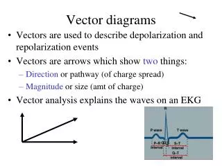
Vector diagrams • Vectors are used to describe depolarization and repolarization events • Vectors are arrows which show two things: • Direction or pathway (of charge spread) • Magnitude or size (amt of charge) • Vector analysis explains the waves on an EKG Q S
EKG is Extracellular Recording • Only looks at the charge on the outside of fibers! • Resting cell: outside positive • Depolarizing cell: outside negative • Repolarizing cell: outside positive • Depolarization: spread of surface neg charge • Repolarization: spread of surface positive charge • Vectors will always be positioned • so that head of vector is in area of positive charge; • tail is in area of negative charge. +++++++++++ ------------------ +++++++++++ ------------------ +++++++++++ ------------------ +++++++++++ ------------------
The Rules of Vectors Analysis • An EKG is a comparison of two vectors • It compares the “heart vector” with the reference “recording lead vector” on the body.
The Rules of Vectors Analysis • If the vectors run parallel (same direction) the pen moves upward from baseline • If the vectors run antiparallel (opposite direction) then the pen moves downward from baseline. • If the vectors are perpendicular, the pen remains on baseline. • If there is no current flow, the pen remains on baseline. • Each lead consists of two electrodes placed on the skin, with a voltmeter between them.
Direction of vector Axis of leads
V analysis of potentials in bipolar leads • Potential • Lead I = ½ • Lead II= equal • Lead III= 1/3
Recording from Lead II II Standard limb lead
- I + - - III II Einthoven’s Triangle Bipolar Limb Leads + +
Atrial depolarization Pen here V II T The heart vector is parallel to the lead, but how can you confirm?
Atrial depolarization - II • Draw a perpendicular line to the lead vector • Draw a line toward from the perpendicular vector toward your cardiac vector +
IV septal depol, from L to R II Anti-parallel! Pen deflects down Draw it!
Lateral walls depol II Draw it!
Depolarization complete; no current flow; pen returns to baseline II
Ventricular Repolarization complete; no current flow; pen on baseline II
Ventricular Repolarization complete; waiting to start all over again II End of one cardiac cycle
Left endo- Cardial surface of septum
repolarization • Surface of heart repolarize first • +ve end of vent. Vector --- towards APEX
How to Plot the Mean Electrical Axis Using Two EKG Leads R wave only = 6mm or .6mv 6mm 6mm RS waves R= 8mm S= -2mm Total = 8-2=6mm or .6mv Mean Electrical Axis
Left axis deviations endomorph- short stature Pregnancy Left ventricle hypertrophy LBBB Right axis deviations Ectomorph- tall /thin Hypertrophy of right ventricle RBBB Mean Axial Shift 76
What exactly is this System? • Which organs are involved? • What is the function of the Lymphatic System? • How does it work?
Jobs of Lymphatic System: Lymphatic System which consists of vessels and organs plays two vital roles in our lives: • The vessels essentially maintain interstitial fluid levels by carrying excess fluids as well as any plasma proteins, back into the CVS. • The organs, house critical immune cells such as lymphocytes which carryout our body defense against infection and disease as well as offer ACQUIRED IMMUNITY .
Lymphatic Characteristics • Lymph – excess tissue fluid carried by lymphatic vessels ( general definition) • Properties of lymphatic vessels • One way system toward the heart • No pump • Lymph moves toward the heart • Milking action of skeletal muscle • Rhythmic contraction of smooth muscle in vessel walls
Composition of Lymph • Lymph is usually a clear, colorless fluid, similar to blood plasma but low is protein • Its composition varies from place to place; • after a meal, for example, lymph draining from the small intestine, takes on a milky appearance, due to lipid content. • Lymph may contain macrophages, viruses, bacteria, cellular debris and even traveling cancer cells.
Lymphatic Vessels • Lymph Capillaries • Walls overlap to form flap-like minivalves • Fluid leaks into lymph capillaries • Capillaries are anchored to connective tissue by filaments • Higher pressure on the inside closes minivalves
Determinants of Lymph Flow • The degree of activity of the lymphatic pump • smooth muscle filaments in lymph vessel cause them to contract • external compression also contributes to lymphatic pumping Figure 16-11; Guyton and Hall
What Type of Vessels Make up the Lymphatic System? • The vessels are called lymphatics. • They are thin-walled and are analogous to veins. • Small lymphatics are similar to capillaries only more porous; Larger vessels are called collecting vessels: both have valves. • 2 large Ducts: Right LYMPHATIC DUCT and THORACIC DUCT (BOTH EMPTY INTO THE RT AND LT SUBCLAVIAN VEINS) • Lymph flows only TO THE HEART (ONE WAY). • This is a low-pressure, pumpless system. Lymph moves via skeletal muscles and pressure changes in thorax during breathing only.
Determinants of Lymph Flow Interstitial fluid hydrostatic pressure Lymph Flow Figure 16-9; Guyton and Hall Figure 16-10; Guyton and Hall
Edema is the excess accumulation of fluids in tissue spaces. This can retard normal exchange of nutrients and metabolites. Filtration of the extracellular fluid exceeds drainage. Anything that causes increased capillary pressure, such as decreased plasma protein, increased capillary permeability or lymphatic blockage, can result in swelling and congestion of the extravascular compartment. EDEMA
Lymph Carries … • Harmful materials that enter lymph vessels • Bacteria • Viruses • Cancer cells • Cell debris