Download
1 / 72
840 likes | 1.54k Views
Dr. Müge Bıçakçıgil Kalaycı. Rheumatoid Arthritis. Second most common form of chronic arthritis Chronic , insidious , autoimmune inflammatory disorder

E N D
Dr. Müge Bıçakçıgil Kalaycı RheumatoidArthritis
Secondmostcommon form of chronicarthritis • Chronic, insidious, autoimmuneinflammatorydisorder • Symmetricpolyarthritisaffectingmainlysmalljoints in thehandandfeet, as well as largerjointssuch as thewristandshoulders RHEUMATOID ARTHRITIS (ra)
Characteristic deformities include subluxations, dislocations, rheumatoid nodules, and joint contractures. • Crippling disease • Shortens survival • quality of life
Twin and family studies demonstrate a heritability of 60%; approximately 30% of genetic risk is attributed to the shared epitope encoded on the human leukocyte antigen molecules. Patients with HLA-DRB1 alleles may have a poorer prognosis
Hormonal and reproductive factors contribute to the female excess and parity, breast feeding, and exogenous hormones are modifiers of risk. Smoking is the strongest known environmental risk factor for RA
The cause of rheumatoid arthritis is unknown Genetic predisposition Enviromental event (such as infection) inappropriate self-directed immune response etIology
envıromental factors • Infectious agents,Immunoregulatory and hormonal irregularities • Immune mediated chronic inflammation • Trigger: Environmental Antigen Genetic (30%) Self Antigen T cell activation Chronic Inflammation Lymphoid cells infiltrate synovium New blood vessels form in synovium Synovial proliferation Joint destruction
Mechanisms of joint damage • Synovial mass stretches joint capsule and ligaments: joint swelling, instability & deformity • Cytokine and proteolytic enzyme rich synovial fluid destroys cartilage joint space narrowing on X-rays • Infiltration of cartilage and later bone by invading synovium (pannus) marginal erosions
Prevalence 0.05-0.15/1000 in developed nations. Disease onset most common at 25-55 years of age Incidence rises with age Women:men 2.5:1 EPİDEMIOLOGY
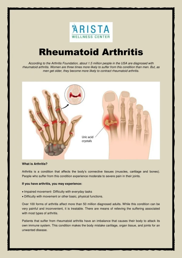
The distribution of involved joint is a critical clue to the underlying diagnosis. RA can effect any of the synovial joints. The disease starts in the MCF, PIP,MTF joints Followed by the wrist, knees, elbows, ankles, hips, and shoulders artIcularmanIfestatIons
Symptoms include pain , swelling, and stiffness Stiffness often predominating in the mornings Early treatment helps limit the number of joints involved RA almost always spares DIP joints
Hand • MCP joints • Synovitis • Ulnar deviation • PIP joints • Synovitis • Swan neck deformity • Boutonniere deformity • Z-deformity of thumb • Tendons • Flexor tenosynovitis • Extensor tenosynovitis • Poor grip: power and pinch
Wrist • Synovitis • Subluxation • Radial deviation • Ankylosis • Carpal tunnel syndrome
Elbow • Synovitis • Flexion contracture • Decreased, painful pronation and supination • Olecranon bursitis • RA nodules
Foot • MTP • Synovitis • Subluxation with hammer toe and metatarsalgia • Toe deviation/overriding • Collapse of medial arch of foot
Ankle/Hindfoot • Ankle • Synovitis • Retrocalcaneal bursitis • Tenosynovitis/rupture • Peroneal tendons • Tibialis posterior • Subtalar arthritis • Reduced and painful movement
Knee • Synovitis • Effusions • Baker’s cyst +/- rupture • Instability/ deformity eg valgus deformity • Flexion contracture
Hip • Arthritis (usually late) • Pain especially on weight bearing • Reduced movement • Trochanteric bursitis SHOULDER • Subacromial bursitis • Rotator cuff tendinitis • Glenohumeral joint arthritis • Acromio-clavicular arthritis
Cervical spine • Involved in 70% patients with longstanding RA • Occipital pain made worse by movement • Subluxation of C1-2 with compression of spinal cord during neck flexion • Significant if >10 mm instability on flexion • Usually slowly developing myelopathy
Other joints • TMJ: reduced mouth opening • Sternoclavicular • Crico-arytenoid • Ossicles of ears
Systemic features; -fatigue, -weight loss, and -low grade fevers (≤ 38 ⁰C) -more common in those patients with rheumatoid factor (seropositive) Extra-articularmanifestations
Approximately one –quarter of patients • In patients who are seropositive for rheumatoid factor. • Nodules may ocur almost anywhere • lungs, heart, and eye, • Most commonly occur subcutaneously on extensor surfaces • particullary forearms , over joints or pressure points Subcutaneousnodules
Firm on examination Usually nontender( unless traumatized) Tought to be triggered by small vessel vasculitis. Subcutaneousnodules
LUNGS • Lungs • Pleurisy • Pleural effusions ( exudate!) • RA nodules single/multiple (Caplan syndrome if huge nodules in coal miners) • Lung fibrosis
RA associated small vessel vasculitis • Digital infarcts • Leukocytoclastic vasculitis • should prompt more aggressive treatment with disease – modifying antirheumatic drugs (DMARDs). • Pyoderma gangrenosum vasculitis
Patients with RA have significantly increased morbidity and mortality from coronary artery disease. • chronic inflammation, • some of medications used, and • sedantary lifestyle may be significant risk factors. cardiacinvolvement
Pericardial effusions • common (detected in up to 50 % of patients by echocardiography) • usually asymptomatic. • Rarely, may result in a fibrinous pericarditis, and constrictive pericarditis • Uncommonly, rheumatoid nodules may occur in the conduction system and cause heart block. cardiacinvolvement
EYES • Secondary Sjögren syndrome • Episcleritis • Scleritis • Scleromalacia perforans
Peripheral nerve entrapment syndromes • Carpal tunel syndrome • Tarsal tunel syndrome • Vasculitis - monoeuritis multiplex • Subluxations at C1-C2 -myelopati. • Rheumatoid nodules in the central nervous system • usually asymptomatic Neurologicmanifestations
The triad of • RA, • Splenomegaly, and • Neutropenia. • In patients with severe , seropositive disease • May be accompanied by hepatomegaly, thrombocytopenia, lympadenopathy and fevers Feltysyndrome
Most patients -do not require specific therapy • Treatment should be focused on severe RA. • Splenectomy -if severe neutropenia exists (<500 cells/µL) a • - recurrent bacterial infections feltysyndrome
Complications • Infections • More susceptible to any infection (RA, steroids, MTX) • ESPECIALLY susceptible to joint infections • Always suspect septic arthritis if sudden increase in symptoms in one joint
Complications • Osteoporosis and fractures • RA • Immobility • Steroids • Amyloidosis • Rare • Longstanding disease • Proteinuria/decreased renal function • Lymphoma