Download
1 / 26
260 likes | 364 Views
This overview explains the fundamental principles of sound as pressure waves characterized by intensity, frequency, and direction. It covers human hearing range, the impact of sound exposure on cochlear hair cells, and common causes of hearing loss such as presbycusis and otosclerosis. The roles of the outer, middle, and inner ear structures in amplifying sound and aiding localization are discussed. Additionally, the functions of hair cells, fluid dynamics within the cochlea, and mechanisms of sound signaling to the brain are explored, highlighting the complexity of auditory processing.

E N D
Sound is pressure waves of alternating compressions and rarefactions. It’s characterized by intensity, frequency, and direction.
• Intensity is in decibels Sound Pressure Level: dB SPL = 20 log [P/P0]. This is relative to an absolute reference. Huge range of sounds are encountered in the world. Human hearing spans 20 – 10,000 Hz, with peak sensitivity at 2 kHz.
• 90dB can produce temporary threshold shift, in which sensitivity is reduced. With severe sound exposure, cochlear hair cells can be killed. They don’t regenerate. Other hearing loss may be iatrogenic (gentamicin). Most common hearing loss is presbycusis, or loss of high frequency hearing with age.
• The pinna collects sound, amplifying it. The ear canal resonates frequencies around 4 kHz and enhances them. These structures help you locate the source of a sound based on frequency signatures.
• The middle ear bones muse overcome the acoustic impedance mismatch of air/water. Water has much higher acoustic impedance, and normally sound going from air to water is mostly reflected. The small area of the stapes footplate and lengths of middle ear bones (lever) increase the pressure wave ~26 fold.
• Tensor tympani and stapedius attenuate transmission through the middle ear when noise is really loud. • A loss of middle ear function is “conductive” hearing loss. It may be caused by otosclerosis, in which bony growth impedes the ear ossicles.
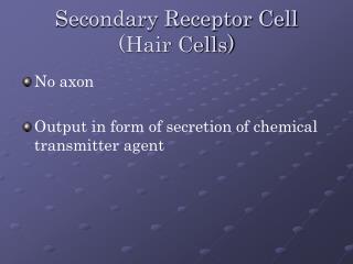
Sensorineural hearing loss may be due to loss of hair cells. They can be differentiated by using bone conduction through the skull to test for an inner ear response.
• External hearing aids may help with both conductive and sensorineural hearing loss. For some conductive hearing loss surgery helps. For severe sensorineural hearing loss, a cochlear implant may be necessary.
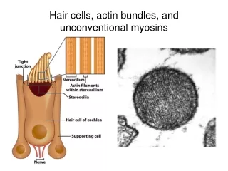
• Movement of stapes footplate at oval window vibrates the scalavestibuli. Scalavestibuli on top, bordered below by Reissner’s membrane, below which is scala media (bounded below by the apical surface of the hair cells). There are inner (1 row) and outer hair cells (3 rows).
At the bottom of the scala media is the cochlear partition (hair cells, basilar membrane and tectorial membrane). Below this is the scala tympani.
• Scala vestibule and tympani have perilymph (low K, high Na), scala media endolymph (high K, low Na). Endolymphis secreted into the scala media by the striavascularis on its outer wall. Hair cells’ apical surfaces are bathed in endolymph, their basolateral surfaces in perilymph.
The apical surfaces have tight junctions to maintain concentration barriers and the +80 mV endolymphatic potential compared to the perilymph. These two forces drive K ions into hair cells at their apical surfaces.
• Meniere’s disease occurs with disrupted endolymphatic fluid circulation and results in sensorineural hearing loss. Vestibular dysfunctions also occurs. • Vertical movement of the basilar and tectorial membranes causes shear between them, pushing stereocilia (actin filled microvilli) on hair cells from side to side.
Hair cells have 1 true cilium called the kinocilium, but it is absent in the adult cochlea. A tip link runs from the top of 1 stereocilium to the side of the adjacent taller one.
• Deflection toward the taller stereocilia results in stretching of the tip link and opening of ion channels at the tips of stereocilia. Deflection in the opposite direction closes channels (at rest 10% are open). The channels are cation-permeant, though the [K] differential and endolymphatic potential lead to a flux of mostly potassium into the hair cell from its apex.
This depolarizes the cell. Sinusoidal deflections of the cochlea result in sinusoidal changes in hair cell membrane potential.
• During a sustained deflection of hair cells, the ion current decays in a form of adaptation. A hypothesis is that motors move the attachment of the tip link to relieve the mechanical activation.
• Hair cell depolarization leads to opening of VG Ca channels that trigger fusion of vesicles containing glutamate. These excite AMPARs on afferent fibers which carry signals to their cell bodies in the spiral ganglion. VG K channels in the basolateral surface of hair cells allow an outward flux of K, and return the cell to its resting potential (hyperpolarizing it really).
• At rest, hair cells release glutamate and afferent neurons have spontaneous activity. So, you can signal by increasing or decreasing the firing rate. An individual cochlear afferent only synapses with a single inner hair cell. At the synapse, the hair cell has a synaptic ribbon to facilitate vesicle release.
• There are 3 outer hair cells for every inner hair cell, but 95% of afferent fibers go to inner hair cells via type I fibers. The contralateral superior olivary complex gives off a crossed olivo-cochlear bundle that synapses massively on outer hair cells.
This pathway is inhibitory, and when it inhibits the outer hair cells it causes a loss of sensitivity and frequency selection in afferent signals from the cochlea. So inhibiting the OHCs alters the response of the IHCs.
• When depolarized, OHCs shorten and when hyperpolarized they lengthen. This electromotility is thought to be carried out by the anion channel prestin, which may be translocated with changes in membrane potential and altering the membrane area. This contributes to the vibration of the cochlear partition and enhances the motion detected by inner hair cells.
• One form of evidence for this idea of active biological motors is that the ear can generate sounds called otoacoustic emissions, which are echoes or distortion products occurring with certain stimulation. Distortion product otoacoustic emissions (DPOAEs) reflect normal activity in the OHCs of the cochlea and may be used to diagnose sensorineural hearing loss.
• Efferent innervations of the cochlea suppresses cochlear sensitivity by inhibiting OHCs. The olivo-cochlear efferents release ACh on the OHCs. The OHCs have a unique nicotinic AChR. This receptor allow a Ca influx and depolarizes the OHC.
But nearby Ca-gated K channels immediately open and hyperpolarize the cell. • Hopefully this unique AChR will provide specific drug targets for the auditory system.