Download
1 / 42
620 likes | 1.18k Views
Antiarrhythmic drugs. Pharma team. LECTURE’S OUTLINE:. Electrophysiology of the heart Arrhythmia: definition, mechanisms, types Drugs :class I, II, III, IV Guide to treat some types of arrhythmia Questions. Physiology of the normal heart. Normal conduction pathway:.

E N D
Antiarrhythmic drugs Pharma team
LECTURE’S OUTLINE: Electrophysiology of the heart Arrhythmia: definition, mechanisms, types Drugs :class I, II, III, IV Guide to treat some types of arrhythmia Questions
Physiology of the normal heart Normal conduction pathway: 1- SA node generates action potential and delivers it to the atria and the AV node Other types of conduction that occurs between myocardial cells: When a cell is depolarized adjacent cell depolarizes along 2- The AV node delivers the impulse to purkinje fibers 3- purkinje fibers conduct the impulse to the ventricles
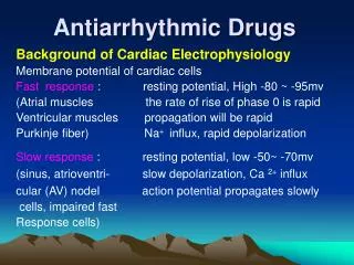
Action potential of the heart: In the SA node and AV node, AP curve consists of 3 phases In the atria, purkinje, and ventricles the AP curve consists of 5 phases
Non-pacemaker action potential Phase 1: partial repolarization Due to rapid efflux of K+ Phase 2: plateu Due to Ca++ influx Phase 0: fast upstroke Due to Na+ influx Phase 3: repolarization Due to K+ efflux Phase 4: resting membrane potential N.B. The slope of phase 0 = conduction velocity Also the peak of phase 0 = Vmax
Pacemaker AP Phase 0: upstroke: Due to Ca++ influx Phase 3: repolarization: Due to K+ efflux Phase 4: pacemaker potential Na influx and K efflux and Ca influx until the cell reaches threshold and then turns into phase 0 Pacemaker cells (automatic cells) have unstable membrane potential so they can generate AP spontaneously
Effective refractory period (ERP) • It is also called absolute refractory period (ARP) : • In this period the cell can’t be excited • Takes place between phase 0 and 3
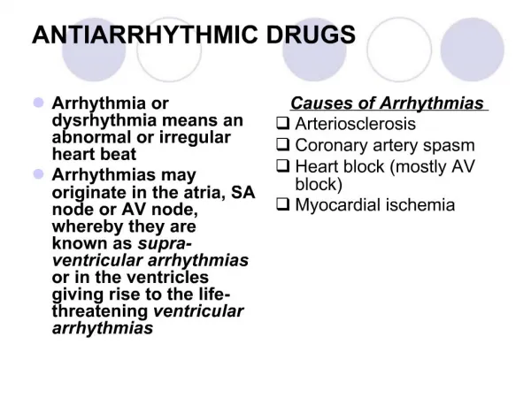
Arrhythmia Arrhythmia /dysrhythmia: abnormality in the site of origin of impulse, rate, or conduction If the arrhythmia arises from atria, SA node, or AV node it is called supraventricular arrhythmia If the arrhythmia arises from the ventricles it is called ventricular arrhythmia
Mechnisms of Arrhythmogenesis Delayed afterdepolarization Early afterdepolarization AP arises from sites other than SA node ↑AP from SA node
1-This pathway is blocked This is when the impulse is not conducted from the atria to the ventricles 3-So the cells here will be reexcited (first by the original pathway and the other from the retrograde) 2-The impulse from this pathway travels in a retrograde fashion (backward)
Abnormal anatomic conduction Here is an accessory pathway in the heart called Bundle of Kent • Present only in small populations • Lead to reexcitation Wolf-Parkinson-White Syndrome (WPW)
Action of drugs In case of abnormal generation: In case of abnormal conduction: Decrease of phase 4 slope (in pacemaker cells) ↑ERP (so the cell won’t be reexcited again) ↓conduction velocity (remember phase 0) Before drug Raises the threshold after phase4
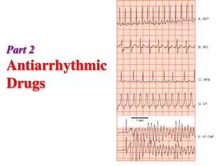
Types of Arrhythmia Supraventricular Arrhythmias • Sinus Tachycardia: high sinus rate of 100-180 beats/min, occurs during exercise or other conditions that lead to increased SA nodal firing rate • Atrial Tachycardia: a series of 3 or more consecutive atrial premature beats occurring at a frequency >100/min • Paroxysmal Atrial Tachycardia (PAT): tachycardia which begins and ends in acute manner • Atrial Flutter: sinus rate of 250-350 beats/min. • Atrial Fibrillation: uncoordinated atrial depolarizations. AV blocks A conduction block within the AV node , occasionally in the bundle of His, that impairs impulse conduction from the atria to the ventricles.
ventricular Arrhythmias • Ventricular Premature Beats (VPBs): caused by ectopic ventricular foci; characterized by widened QRS. • Ventricular Tachycardia (VT): high ventricular rate caused by abnormal ventricular automaticity or by intraventricular reentry; can be sustained or non-sustained (paroxysmal); characterized by widened QRS; rates of 100 to 200 beats/min; life-threatening. • Ventricular Flutter- ventricular depolarizations >200/min. • Ventricular Fibrillation- uncoordinated ventricular depolarizations
Pharmacologic Rationale & Goals • The ultimate goal of antiarrhythmic drug therapy: • Restore normal sinus rhythm and conduction • Prevent more serious and possibly lethal arrhythmias from occurring. • Antiarrhythmic drugs are used to: • decrease conduction velocity • change the duration of the effective refractory period (ERP) • suppress abnormal automaticity
Antyarrhythmic drugs • Most antiarrhythmic drugs are pro-arrhythmic (promote arrhythmia) • They are classified according to Vaughan William into four classes according to their effects on the cardiac action potential
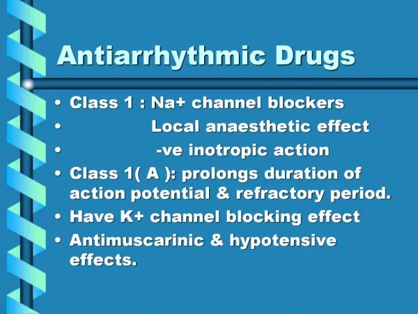
Class I drugs Have moderate K+ channel blockade They ↓ conduction velocity in non-nodal tissues (atria, ventricles, and purkinje fibers) They act on open Na+ channels or inactivated only So they are used when many Na+ channels are opened or inactivated (in tachycardia only) because in normal rhythm the channels will be at rest state so the drugs won’t work
Mechanism of action • Slowing of the rate of rise in phase 0 ↓conduction velocity • ↓of Vmaxof the cardiac action potential • They prolong muscle action potential & ventricular (ERP) • They ↓ the slope of Phase 4 spontaneous depolarization (SA node) decrease enhanced normal automaticity They make the slope more horizontal
Class IA Drugs • They possess intermediate rate of association and dissociation (moderate effect) with sodium channels. Pharmacokinetics: Procainamide metabolized into N-acetylprocainamide (NAPA) (active class III) which is cleared by the kidney (avoid in renal failure)
Class IA Drugs Uses • Supraventricular and ventricular arrhythmias • Quinidine is rarely used for supraventricular arrhythmias • Oral quinidine/procainamide are used with class III drugs in refractory ventricular tachycardia patients with implantable defibrillator • IV procainamide used for hemodynamically stable ventricular tachycardia • IV procainamide is used for acute conversion of atrial fibrillation including Wolff-Parkinson-White Syndrome (WPWS) defibrillator
Class IA Drugs Toxicity • Systemic lupus erythromatosus (SLE)-like symptoms: arthralgia, fever, pleural-pericardial inflammation. • Symptoms are dose and time dependent • Common in patients with slow hepatic acetylation
Notes: Torsades de pointes: twisting of the point . Type of tachycardia that gives special characteristics on ECG At large dosesofquinidinecinchonismoccurs:blurred vision, tinnitus, headache, psychosis and gastrointestinal upset Digoxin is administered before quinidine to prevent the conversion of atrial fibrillation or flutter into paradoxical ventricular tachycardia
Class IB Drugs • They shorten Phase 3 repolarization • ↓ the duration of the cardiac action potential • They suppress arrhythmias caused by abnormal automaticity • They show rapid association & dissociation(weak effect) with Na+ channels with appreciable degree of use-dependence • No effect on conduction velocity
Agents of Class IB Lidocaine • Used IV because of extensive 1st pass metabolism • Lidocaine is the drug of choice in emergency treatment of ventricular arrhythmias • Has CNS effects: drowsiness, numbness, convulstion, and nystagmus Mexiletine • These are the oral analogs of lidocaine • Mexiletine is used for chronic treatment of ventricular arrhythmias associated with previous myocardial infarction Adverse effects: 1- neurological effects 2- negative inotropic activity • Uses • They are used in the treatment of ventricular arrhythmias arising during myocardial ischemia or due to digoxin toxicity • They have little effect on atrial or AV junction arrhythmias (because they don’t act on conduction velocity)
Class IC Drugs • They markedly slow Phase 0 fast depolarization • They markedly slow conduction in the myocardial tissue • They possess slow rate of association and dissociation (strong effect) with sodium channels • They only have minor effects on the duration of action potential and refractoriness • They reduce automaticity by increasing the threshold potential rather than decreasing the slope of Phase 4 spontaneous depolarization.
Uses: • Refractory ventricular arrhythmias. • Flecainide is a particularly potent suppressant of premature ventricular contractions (beats) Toxicity and Cautions for Class IC Drugs: • They are severe proarrhythmogenic drugs causing: • severe worsening of a preexisting arrhythmia • de novo occurrence of life-threatening ventricular tachycardia • In patients with frequent premature ventricular contraction (PVC) following MI, flecainide increased mortality compared to placebo. Notice: Class 1C drugs are particularly of low safety and have shown even increase mortality when used chronically after MI
Compare between class IA, IB, and IC drugs as regards effect on Na+ channel & ERP • Sodium channel blockade: IC > IA > IB • Increasing the ERP:IA>IC>IB (lowered) Because of K+ blockade
Class II ANTIARRHYTHMIC DRUGS(β-adrenergic blockers) Uses • Treatment of increased sympathetic activity-induced arrhythmias such as stress- and exercise-induced arrhythmias • Atrial flutter and fibrillation. • AV nodal tachycardia. • Reduce mortality in post-myocardial infarction patients • Protection against sudden cardiac death Mechanism of action • Negative inotropic and chronotropic action. • Prolong AV conduction (delay) • Diminish phase 4 depolarization suppressing automaticity(of ectopic focus)
Class II ANTIARRHYTHMIC DRUGS • Propranolol (nonselective): was proved to reduce the incidence of sudden arrhythmatic death after myocardial infarction • Metoprolol • reduce the risk of bronchospasm • Esmolol: • Esmolol is a very short-acting β1-adrenergic blocker that is used by intravenous route in acute arrhythmias occurring during surgery or emergencies selective
Class III ANTIARRHYTHMIC DRUGSK+ blockers • Prolongation of phase 3 repolarizationwithout altering phase 0 upstroke or the resting membrane potential • They prolong both the duration of the action potential and ERP • Their mechanism of action is still not clear but it is thought that they block potassium channels
Uses: • Ventricular arrhythmias, especially ventricular fibrillation or tachycardia • Supra-ventricular tachycardia • Amiodarone usage is limited due to its wide range of side effects
Sotalol (Sotacor) • Sotalol also prolongs the duration of action potential and refractoriness in all cardiac tissues (by action of K+ blockade) • Sotalol suppresses Phase 4 spontaneous depolarization and possibly producing severe sinus bradycardia (by β blockade action) • The β-adrenergic blockade combined with prolonged action potential duration may be of special efficacy in prevention of sustained ventricular tachycardia • It may induce the polymorphic torsades de pointes ventricular tachycardia (because it increases ERP) • Ibutilide • Used in atrial fibrillation or flutter • IV administration • May lead to torsade de pointes • Only drug in class three that possess pure K+ blockade
Amiodarone (Cordarone) • Amiodarone is a drug of multiple actions and is still not well understood • It is extensively taken up by tissues, especially fatty tissues (extensive distribution) • t1/2 = 60 days • Potent P450 inhibitor • Amiodarone antiarrhythmic effect is complex comprising class I, II, III, and IV actions • Dominant effect: Prolongation of action potential duration and refractoriness • It slows cardiac conduction, works as Ca2+ channel blocker, and as a weak β-adrenergic blocker Toxicity • Most common include GI intolerance, tremors, ataxia, dizziness, and hyper-or hypothyrodism • Corneal microdeposits may be accompanied with disturbed night vision • Others: liver toxicity, photosensitivity, gray facial discoloration, neuropathy, muscle weakness, and weight loss • The most dangerous side effect is pulmonary fibrosiswhich occurs in 2-5% of the patients
Class IV ANTIARRHYTHMIC DRUGS (Calcium Channel Blockers) • Calcium channel blockers decrease inward Ca2+ currents resulting in a decrease of phase 4 spontaneous depolarization (SA node) • They slow conductance in Ca2+ current-dependent tissues like AV node. • Examples: verapamil & diltiazem Because they act on the heart only and not on blood vessels. • Dihydropyridine family are not used because they only act on blood vessels
Mechanism of action • They bind only to depolarized (open) channels prevention of repolarization • They prolong ERP of AV node ↓conduction of impulses from the atria to the ventricles So they act only in cases of arrhythmia because many Ca2+ channels are depolarized while in normal rhythm many of them are at rest Uses • More effective in treatment of atrial than ventricular arrhythmias. • Treatment of supra-ventricular tachycardia preventing the occurrence of ventricular arrhythmias • Treatment of atrial flutter and fibrillation
contraindication • Contraindicated in patients with pre-existing depressed heart function because of their negative inotropic activity Adverse effects • Cause bradycardia, and asystole especially when given in combination with β-adrenergic blockers
Miscellaneous Antiarrhythmic Drugs Adenosine • Adenosine activatesA1-purinergic receptors decreasing the SA nodal firing and automaticity, reducing conduction velocity, prolonging effective refractory period, and depressing AV nodal conductivity • It is the drug of choice in the treatment of paroxysmal supra-ventricular tachycardia • It is used only by slow intravenous bolus • It only has a low-profile toxicity (lead to bronchospasm) being extremly short acting for 15 seconds only
Treatment of atrial flutter/fibrillation 1st: Reduce thrombus formation by using anticoagulant warfarin • 2nd: Prevent the arrhythmia from converting to ventricular arrhythmia: • First choice: class II drugs: • After MI or surgery • Avoid in case of heart failure • Second choice: class IV • Third choice: digoxin • Only in heart failure of left ventricular dysfunction 3rd: Conversion of the arrhythmia into normal sinus rhythm: Class III: IV ibutilide, IV/oral amiodarone, or oral sotalol Class IA: Oral quinidine + digoxin (or any drug from the 2nd step) Class IC: Oral propaphenone or IV/oral flecainide Use direct current in case of unstable hemodynamic patient
Treatment of ventricular arrhythmia Premature ventricular beat (PVB) • First choice: class II • IV followed by oral • Early after MI • Second choice: amiodarone Avoid using class IC after MI ↑ mortality Treatment of ventricular tachycardia • First choice: Lidocaine IV • Repeat injection • Second choice: procainamide IV • Adjust the dose in case of renal failure • Third choice: class III drugs • Especially amiodarone and sotalol
تجميعات: (IA, IC, class III) torsades de pointes. Classes II and IV bradycardia (don’t combine the two) In atrial flutter use (1st↓impulses from atria to ventricular to prevent ventricular tachycardia) Class II Class IV Digoxin. (على الترتيب) 2nd convert atrial flutter to normal sinus rhythm use: Ibutilide Sotalol IA or IC. (على الترتيب) If you use quinidine combine it with digoxin or β blocker (because of its anti muscarinic effect) Avoid IC in myocardial infarction because it ↑ mortality
questions 1- In ventricular tachycardia and stable hemodynamic which drug to be used? A- propranolol B- procainamide C- quinidine D- verapamil 2- Mr.Green devloped an arrhythmia and was treated. A month later, he has arthralgia, fever, pleural inflammation. What was the treatment of arrhythmia? A- esmolol B- class III C- procainamide D- propafenone 3- Cinchonism occurs with digoxin A- pulmonary fibrosis diltiazem B- bradycardia amiodarone (F)