Download
1 / 73
800 likes | 1.23k Views
Antiarrhythmic Drugs. Background of Cardiac Electrophysiology Membrane potential of cardiac cells Fast response : resting potential, High -80 ~ -95mv (Atrial muscles the rate of rise of phase 0 is rapid Ventricular muscles propagation will be rapid

E N D
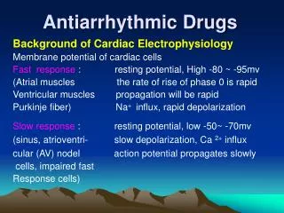
Antiarrhythmic Drugs Background of Cardiac Electrophysiology Membrane potential of cardiac cells Fast response : resting potential, High -80 ~ -95mv (Atrial muscles the rate of rise of phase 0 is rapid Ventricular muscles propagation will be rapid Purkinje fiber) Na+ influx, rapid depolarization Slow response : resting potential, low -50~ -70mv (sinus, atrioventri- slow depolarization, Ca 2+ influx cular (AV) nodel action potential propagates slowly cells, impaired fast Response cells)
1 2 0mV 0 3 100ms 4 -85mV Na+ Ca2+ Na+ Na+ Outside Membrance intside K+ Ca2+ K+,Cl-Channel currents Pump Exchanger Phase 0 : depolarization Phase 1,2,3 : repolarization Phase 4 : diastolic voltage time course 0 ~ 3 : action potential duration APD Fast response
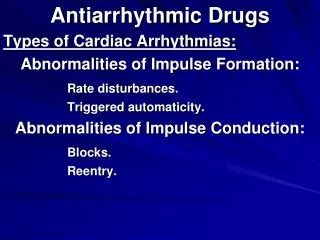
Excitability: relationship between threshold potentialand restingpotential level • Automaticity: • Conductivity: conductive rate is dependent on membrane responsiveness Membrane responsiveness: relationship between Vmax of phase 0 and membrane potential level 4. Effective refractory period, ERP The time between phase 0 and sufficient recovery of sodium channels in phase 3 to permit a propagated response to external stimulus is the “refractory period” .
二 Mechanisms of arrhythmias 1. Disturbances of impulse formation (冲动形成障碍) ① The changes of normal autonomic mechanism Change of pacemaker current (cell) of diastolic autonomic depolarization can cause autonomic alteration such as : mental stress (tension) drug toxicity fever excitation
② formation of abnormal autonomic mechanism non-autonomic cell: atrial muscles ventricular muscles autonomic cell resting potential : -60mv abnormal autonomy repetitive impulse arrhythmias
2. triggered activity (触发活动) and Afterdepolarization (后除极) A Early afterdepolarization (EAD,早后除极) Occur in phase 2, 3 , low potassium, Ca 2+ inward E.A. is secondary depolarization that occur before repolarization is complete. secondary depolarization commences at membrane potentials close to those present during the plateau of the action potential B delayed afterdepolarization ( DAD, 迟后除极) Occur in phase 4, Ca 2+ overload in cell, Na + inward. D.A. is a secondary depolarization that occurs early in diastole, that is, after full repolarization has been achieved.
3. Disturbances of impulse conduction (冲动传导障碍) A causing partial and complete block B reentry---fibrillation心室纤颤 and flutter心室扑动 tachycardia extra beats ( extrasystoles) formation unidirectional block of cardial tissue of reentry circuiting tract shortening the effective refractory period
Reentry circuit established 1 forward impulse obstructed and extinguished 2 decremental conduction(递减传导) and unidirectional block (单向阻滞) of antegrade(顺行) impulse 3 retrograde(逆行) impulse conducted across depressed region 4 reentry circuit established Arrhythmia may be manifest as one or a few extra beats or as a sustained tachycardia
Reentry(折返) : circus movement one impulse reenters and excites areas of the heart more than once
Mechanism of the reentry 正常心肌 单向传导阻滞 折返形成
4. Genetics/gene mutation • Long Q-T SYNDROME, LQTS • 3 mutation genes: SCN5A /chromosome 3: coding sodium channel in myocardia; HERG/ chromosome 7: coding Ikr potassium channel (内向整流钾通道); KVLQT1/ chromosome 11: coding Iks potassium channel (延迟整流钾通道); 5. Disturbances of Potassium channels
Classification of Antiarrhythmic Drugs Antiarrhythmic agents are divided into FOUR classes Assignment to the respective classes is made on the basis of drug-induced alterations in ion channel function and cardiac electrophysiologic properties The classification, while helpful, is not absolute and overlapping properties exist among the many drugs.
A. Antiarrhythmic drugs can depress Na + inward of non-autonomic cell in phase 4 or depress Ca 2+ inward of autonomic cell in phase 4 depress automaticity • B. Antiarrhythmic drugs can accelerate K+ outward of phase 3, increase maximum diastolic potential (more negative ) increase voltage difference between maximum diastolic potential and threshold potential depress automaticity Question: How about the conduction?
Classes of antiarrhythmic agents 1. sodium channel blocking drugs. 2. blockade of sympathetic autonomic effects in the heart 3. prolongation of the ERP and APD 4. calcium channel blockade
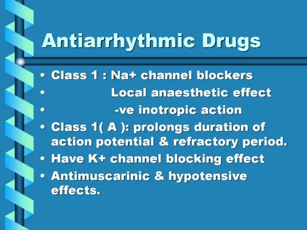
Classification of Antiarrhythmic Drugs Classification I:sodium channel blocking drugs • Class Ia-Characteristics Meddle level sodium channel block,weak level potassium channel block, and weak level calcium channel block in high concentration • Slow the rate of rise of the membrane action potential (Phase 0; dV/dt ) • Slow conduction velocity (PR; QRS) • Prolong refractoriness (QT) • Examples - • Quinidine* • Procainamide • Disopyramide
Classification of Antiarrhythmic Drugs • Class Ib -Characteristics Weak level sodium channel block,and potassium channel open • Limited effect on dV/dt of Phase 0 • Slight slowing of conduction velocity • No change or a decrease in refractory period • Examples • Lidocaine* • Tocainide • Mexiletine • Moricizine ?
Classification of Antiarrhythmic Drugs Class Ic- Characteristics Strong level sodium channel block,and weak level calcium channel block • Marked slowing of conduction velocity (prolongs PR and QRS) • No change in refractoriness or repolarization • Examples • Flecainide* • Propafenone (also Class II) • Moricizine (also Class Ib) • Encainide (discontinued)
Classification of Antiarrhythmic Drugs • Class II-Characteristics • Produce beta-adrenergic receptor blockade (prolongs PR; slows heart rate) • Decrease in refractory period duration (decrease in QT) • Examples • Propranolol* • Acebutolol • Esmolol • Sotalol (also Class III)
Classification of Antiarrhythmic Drugs • Class III-Characteristics potassium channel block • Prolong the action potential duration • Increase the refractory period (increase in the QT) • Examples • Amiodarone (also some Class Ia,II,III,&IV) • Bretylium • Sotalol (also Class II) • Ibutilide*
Classification of Antiarrhythmic Drugs • Class IV-Characteristics • Blockade of calcium entry via slow inward channel (prolong the PR interval) • Examples • Verapamil • Diltiazem
Other Miscellaneous Agents • Adenosine • Depresses sinus node automaticity • Depresses atrioventricular node conduction • Uses • Acute termination of AV nodal tachycardia • Acute termination of AV nodal reentrant tachycardia
Other Miscellaneous Agents • Digitalis (Digoxin) • Prolongs atrioventricular nodal conduction time and increases functional refractory period - directly and indirectly (increase in vagal cholinergic tone) • Slows sinus rate when ventricular function is impaired by virtue of its direct positive inotropic effect (withdrawal of sympathetic tone) • Uses: • Atrial fibrillation or flutter - primarily to control the ventricular rate • AV nodal reentrant tachycardia
Class Ia Antiarrhythmic Agents • Quinidine (奎尼丁) • Procainamide (普鲁卡因胺) • Disopyramide(丙吡胺)
Quinidine (奎尼丁) Electrophysiology inhibit Na+ inward, inhibit K+ outward,inhibit calcium inward in high concentration, depress slope phase 4 diastolic depolarization Pharmacologic action 1 Quinidine depresses pacemaker rate, especially that of ectopic pacemakers ( abnormal automaticity) depress automaticity of atrial, ventricular muscles, Purkinje, and sinoatrial nodes
2 Quinidine also lengthens the action potential duration ( APD) and effective refractory period (ERP) depresses phase 3 K+ outward, slow repolarization lengthens the APD, and ERP eliminates reentry impulses.
3 Negative conduction blocks sodium channel, depresses Na + inward, reduces depolarization rate of phase 0, inhibits conduction responsiveness of membrane declines. inhibits vagal activity, increases conduction of atrioventri-cular (AV) nodes, slow conduction of atrial muscles( reduce atrial bates) and increase the ventricular bates (ventricular fibrillation心室纤颤 and flutter心室扑动)
treating atrial fibrillation and flutter: combination with cardiac lycosides (digoxin), inhibiting conduction of AV node to prevent the ventricular bates. • unidirectional block bidirectional block by abolished reentry impulse. 4 Electrocardiogram (ECG) • QT interval is prolonged • QRS wave is widened
Pharmacokinetics absorption : orally, rapid, in gastrointestinal tract binding protein : 80% bioavailability (F) :72%~87% Vd : 2~3 L/kg metabolism : in liver excretion : 20% unchanged in the urine t ½ 5~7 hours urinary excretion is enhanced in acid urine t ½ may congestive heart failure be longer hepatic or renal diseases older patients
Clinical uses 1 acute and chronic ventricular and supraventricular arrhythmias 2 most common indications: atrial fibrillation and flutter combination with digoxin 3Qinidine can increase blood concentration and untoward reaction of digoxin.
Toxicity • 1 Toxic dosage • depresses conduction of sinoatrial, atrial-ventricular nodes and Purkinje, cause conductive block of atrioventricle and intraventricle. • severe toxication: automaticity of Purkinje can be enhanced, • cause ventricular tachycardia and ventricular fibrillation (may be fatal) iv NaHCO3, K+ inward, K+ in blood is decreased, toxicity is decreased.
2 hypotention Quinidine can block α- receptor, blood vessels relaxation ( vasodilation), inhibit myocardial concentrating force 3 thromboembolism patient with atrial fibrillation easy to occur. 4 cinchonism 金鸡钠中毒 headache, dizziness, tinnitus(耳鸣),confused vision(视力模糊), double vision(复视), gastrointestinal discomfort, fainting晕厥, psycholeptic episodes 精神失常 ( psychataxia mentation), confusion(神志不清) 5 others diarrhea, nausea and vomiting, thrombocytopenia(血小板减少症), bleeding
4 cinchonism 金鸡钠中毒 • headache, dizziness, tinnitus(耳鸣),confused vision(视力模糊),double vision(复视),gastrointestinal discomfort, • fainting晕厥, psycholeptic episodes 精神失常 • ( psychataxia mentation) ,confusion(神志不清) • 5 others diarrhea , nausea and vomiting, thrombocytopenia(血小板减少症), bleeding
Classification of Antiarrhythmic Drugs • Class Ib -Characteristics weak inhibit Na + inward enhance K+ outward depress slope phase 4 diastolic • Examples • Lidocaine* (利多卡因) • Phenytoin sodium(苯妥英钠)
Lidocaine/利多卡因 • Action 1. depressing automaticity (therapeutic dose) lidocaine can suppress automaticity of Purkinje fibers, because of: weak inhibit Na + inward enhance K+ outward depress slope phase 4 diastolic depolarization
2. duration of the action potential (APD) and effective refractory period (ERP) in Purkinje fibers and ventricular muscle: the drug can decrease (shorten) APD and ERP, but decreased APD > decreased ERP. ERP is prolonged relatively APD is shortened Repolarization is rapid and complete, velocity of phase 0 depolarization can be quickened
3. conductivity in condition of ischemic Purkinje fibers of myocardial infarction region the drug can inhibit Na+ inward decrease conduction prevent occur of reentry (from unidirectional block changes to bidirectional block ) in condition of extracellular low K+ or partial depolarization of myocardial tissues the drug can enhance phase 3 K+ outward causing hyperpolarezition, improving conduction abolishing ventricular reentry (reducing unidirectional block)
1 2 0mV 0 3 4 -85mV ERP ADP
Pharmacokinetics 1 very extensive first- pass hepatic metabolism ,only 3% of orally administered lidocaine appears in plasma the concentration in plasma is low Thus, lidocaine must be given parenterally. im. iv. 2 protein binding rate is about 70% 3 t ½ is about 100 min 5~7h Css
Therapeutic use • 1 ventricular arrhythmias ventricular tachycardia and fibrillation • 2 ventricular arrhythmias caused by acute myocardial infarction • 3 open-heart surgery and digitalis toxication
Untoward effects • 1 CNS lightheadedness 头晕 headache paresthesias 感觉异常 (often perioral 口周的) muscle twitching 抽搐 convulsion 惊厥 slurred speech 少言少语 hearing disturbances 听力失调 respiratory arrest 呼吸停止 • 2 hypotension (partly by depressing myocardial contractility) sinoatrial nodal standstill 窦性停搏 impaired conduction • Contraindication ⅡⅢ---atrioventricular conduction block
Phenytoin sodium苯妥英钠 • The drug for the treatment of seizures(癫痫病发作) • Clinical usefulness for ventricular arrhythmias,especially those associated with digitalis toxicity .
Action 1. Automaticity Hastening k+ outward Decreasing the slope of normal phase-4 depolarization in Purkinje fibers (increasing maximal diastolic potential.) automaticity of abolishing delayed Purkinje fibers afterdepolarization caused by digitalis toxicity.
2. APD and ERP in ventricular muscle and Purkinje fibers APD and ERP are shortened, but shortened APD >shortened EPR, so, EPR is rolonged relatively The drug substanitially decrease the APD. Complete repolarization. Level of membrane potential ( negtive potential) Amplitude of action potential Conduction velocity Abolishing reentry.
3.Responsiveness and conduction. Increasing phase-0 depolarization rate of atrial muscle, atrioventricular node, Purkinje fibers of digitalis toxicity. Improving conduction.
Therapeutic uses: • 1.Ventricular arrhythmias. • 2.Paroxysmal atrial flutter or fibrillation. • 3.Supraventricular arrhythmias (tachycardia) • 4.Ventricular arrhythmias caused by acute myocardial infarction, open-heart surgery and digitalis toxication