Download
1 / 93
950 likes | 1.22k Views
Evaluation and Management of Acute Pericarditis. DR.GHYATH AL AGHA CARDIOLOGIST. INTRODUCTION.

E N D
Evaluation and Management of Acute Pericarditis DR.GHYATHAL AGHA CARDIOLOGIST
The pericardium is a fibroelastic sac made up of visceral and parietal layers separated by a (potential) space, the pericardial cavity. In healthy individuals, the pericardial cavity contains 15 to 50 mL of an ultrafiltrate of plasma.
Acute pericarditis is a common disorder in several clinical settings, and may be the first manifestation of an underlying systemic disease..
The pericardium may be involved in a large number of systemic disorders or may be diseased as an isolated process
Idiopathic In most case series, the majority of patients are not found to have an identifiable cause of pericardial disease. Frequently such cases are presumed to have a viral or autoimmune etiology.
Infections Viral Coxsackievirus, echovirus, adenovirus, EBV, CMV, influenza, varicella, rubella, HIV, hepatitis B, mumps, parvovirus B19, vaccina (smallpox vaccination)
Bacterial Staphylococcus Streptococcus pneumococcus Haemophilus Neisseria (gonorrhoeae or meningitidis) Chlamydia (psittaci or trachomatis) Legionella Tuberculosis Salmonella Mycoplasma Lyme disease
Fungal Histoplasmosis, aspergillosis, blastomycosis, coccidiodomycosis, actinomycosis, nocardia, candida
Parasitic Echinococcus, amebiasis, toxoplasmosis
Neoplasm Metastatic - Lung or breast cancer, Hodgkin's disease, leukemia, melanoma Primary - Rhabdomyosarcoma, teratoma, fibroma, lipoma, leiomyoma, angioma Paraneoplastic
Cardiac A.Early infarction pericarditis B. Late postcardiac injury (Dressler's syndrome ) C. Myocarditis
Trauma A. Blunt B. Penetrating C. Iatrogenic - Catheter and pacemaker perforations, cardiopulmonary resuscitation, post-thoracic surgery
Autoimmune A. Rheumatic diseases - including lupus, rheumatoid arthritis, vasculitis, scleroderma, mixed connective tissue disease B. Other - Wegener's granulomatosis, polyarteritisnodosa, sarcoidosis, inflammatory bowel disease (Crohn's, ulcerative colitis), Whipple's, giant cell arteritis, Behcet's disease
Drugs A. Procainamide, isoniazid, or hydralazine as part of drug-induced lupus B. Other - cromolyn sodium, dantrolene, methysergide, anticoagulants, thrombolytics, phenytoin, penicillin, ,mesalazine ,cyclosporin ,phenylbutazone, doxorubicin
Metabolic A. Hypothyroidism B. Uremia
DIAGNOSTIC CRITERIA AND CLINICAL PRESENTATION of ACUTE PERICARDITIS
Acute Pericarditis at least 2 criteria of 4 should be present
1. Typical chest pain 2. Pericardial friction rub 3. Suggestive ECG changes (typically widespread ST segment elevation) 4. New or worsening pericardial effusion (Pericardial effusion confirms the clinical diagnosis but its absence does not exclude it.)
Chest pain The chest pain of acute pericarditis is typically fairly sudden in onset and occurs over the anterior chest. It is often pleuritic in nature, being sharp and exacerbated by inspiration. However, dull, oppressive pain, which is difficult to distinguish from that of myocardial ischemia, can occur. The pain may decrease in intensity when the patient sits up and leans forward
Pericardial friction rub A pericardial friction rub is highly specific for acute pericarditis. The sensitivity is variable, varying in part with the frequency of auscultation since rubs tend to vary in intensity and can come and go over a period of hours. Pericardial rubs may be easier to hear in patients without a pericardial effusion, but this finding is not universal and is not well-documented. In a report of 100 patients with acute pericarditis, a pericardial rub was present in 34 of 40 (85 percent) without an effusion, a prevalence considerably higher than in some other series.
Pericardial friction rubs are said to be generated by friction of the two inflamed layers of the pericardium, but even a large pericardial effusion does not necessarily prevent a friction rub. Thus, this commonly offered explanation for its mechanism may be an oversimplification.
Pericardial friction rubs occur during the maximal movement of the heart within its pericardial sac. Thus, the classic friction rub consists of three phases, corresponding to movement of the heart during atrial systole (which is not heard in patients with atrial fibrillation), ventricular systole, and in the rapid filling phase of early ventricular diastole.
However, some rubs are present only during one (one component) or two phases (two components) of the cardiac cycle
Pericardial rubs have a superficial scratchy or squeaking sound that is best heard with the diaphragm of the stethoscope. They can be localized or widespread, but are usually best heard over the left sternal border..
The intensity of the rub frequently increases after application of firm pressure with the diaphragm, during suspended respiration, and with the patient leaning forward or resting on elbows and knees. This last maneuver is designed to increase contact between visceral and parietal pericardium, but is seldom used in practice since it is cumbersome for the patient
Electrocardiogram The electrocardiogram in acute pericarditis evolves through four stages
Stage 1 Seen in the first hours to days Characterized by diffuse ST elevation (typically concave up) with reciprocal ST depression in leads aVR and V1 There is also an atrial current of injury, reflected by elevation of the PR segment in lead aVR and depression of the PR segment in other limb leadsand in the left chest leads, primarily V5 and V6. Thus, the PR and ST segments typically change in opposite directions, although the PR deviations, which are highly specific although not sensitive, are frequently overlooked.
Stage 2 Characterized by normalization of the ST and PR segments.
Stage 3 Characterized by the development of diffuse T wave inversions, generally after the ST segments have become isoelectric. However, this stage is not seen in some patients.
Stage 4 The ECG may become normal ORthe T wave inversions may persist indefinitely
Treatment can accelerate or alter ECG progression. The duration of the ECG changes in pericarditis also depends upon its cause and the extent of the associated myocardial damage
Atypical ECG changes Atypical ECG such as localized ST-elevation and T-wave inversion before ST-segment normalization occur in a minority of patients with acute pericarditis without myocardial involvement. These changes can simulate changes found in acute coronary syndrome.
Laboratory signs of inflammation Common in patients with acute pericarditis. These include elevations in the white blood cell count, erythrocyte sedimentation rate, and serum C-reactive protein concentration.
Cardiac Biomarkers Acute pericarditis may be associated with increases in serum biomarkers for myocardial injury such as elevations in serum cardiac troponin I (cTnI) with or without increased MB fraction of creatinekinase (CK-MB). Such patients should be considered to have myopericarditis.
1. Definite diagnosis of acute pericarditis, PLUS 2. Suggestive symptoms (dyspnea, palpitations, or chest pain) andECG abnormalities beyond normal variants, not documented previously (ST/T abnormalities, supraventricular or ventricular tachycardia or frequent ectopy, atrioventricular block), OR focal or diffuse depressed LV function of uncertain age by an imaging study 3. Absence of evidence of any other cause 4. One of the following features: evidence of elevated cardiac enzymes (creatinekinase-MB fraction, or troponin I or T), OR new onset of focal or diffuse depressed LV function by an imaging study, OR abnormal imaging consistent with myocarditis (MRI with gadolinium, gallium-67 scanning, anti-myosin antibody scanning)
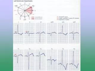
Causes of ST segment elevation Myocardial ischemia or infarction Noninfarction, transmural ischemia (Prinzmetal's angina pattern or acute takotsubocardiomyopathy) Acute myocardial infarction (MI) Post-MI (ventricular aneurysm pattern) Previous MI with recurrent ischemia in the same area Acute pericarditis Normal "early repolarization variants" Left ventricular hypertrophy or left bundle branch block (only V1-V2 or V3) Other Myocarditis (may look like myocardial infarction or pericarditis) Brugada patterns (V1-V3 with right bundle branch block-appearing morphology) Myocardial tumor Myocardial trauma Hyperkalemia (only leads V1 and V2) Hypothermia (J wave/Osborn wave)
Distinction from acute myocardial infarction The electrocardiographic changes in acute pericarditis differ from those in acute ST elevation MI (STEMI) in the following ways These distinctions assume that the pericarditis does not occur during or soon after an acute MI
ACUTE PERICARDITIS STEMI The ST segment elevation begins at the J point, rarely exceeds 5 mm, and usually retains its normal concavity. In other cases, ST segment rises obliquely in a straight line Although similar patterns can occur with STEMI, the typical finding is convex (dome-shaped) ST elevation,a pattern not characteristic of acute pericarditis, that may be more than 5 mm in height
ACUTE PERICARDITIS STEMI The pericardium envelops the heart, and the ST-T changes are therefore more generalized, being present in most of the chest leads as well as leads I, aVL, II, III, and aVF ST segment elevation in the precordial leads is most commonly seen in V5 and V6, and in decreasing frequency from V4 to V1 in precordial leads. In the limb leads, it is often more evident in leads I and II than in leads III, aVF, and aVL. ST segment elevations are characteristically limited to either the anterolateral leads (I, aVL, V1 to V6) or the inferior (II, III, aVF) leads because of the localized area of the infarct
ACUTE PERICARDITIS STEMI Reciprocal ST segment changes, are not seen except in aVR and V1. Often associated with reciprocal ST segment changes.
ACUTE PERICARDITIS STEMI ST segment elevation and T wave inversions do not generally occur simultaneously ST segment elevation and T wave inversions are commonly seen
ACUTE PERICARDITIS STEMI The myocardial injury is more diffuse in and different areas of myocardium reflect different stages in the pattern of repolarizationabnormality.As a result, varying degrees of T wave inversion ORST segment elevation can be present concurrently in different leads Leads facing the infarcted area tend to show the same stage of ST-T evolution