Download
1 / 20
230 likes | 648 Views
Review of the hydrodynamic model of CSF flow including interpretations by Bulat. Ian Gopal Gould UIC – LPPD October 20, 2010. Recent insights into a new hydrodynamics of the cerebrospinal fluid Marin Bulat, Marijan Kalrica, Brain Research Review, August 2010.

E N D
Review of the hydrodynamic model of CSF flowincluding interpretations by Bulat Ian Gopal Gould UIC – LPPD October 20, 2010
Recent insights into a new hydrodynamics of the cerebrospinal fluidMarin Bulat, Marijan Kalrica, Brain Research Review, August 2010 • A consistent author in hydrodynamics since 1970’s – this review summarizes 40 years in the field. • A radical claim: that absorption of CSF into the venous sinuses via physiological pressure is of minor importance due to their minute surface area compared to the huge absorptive area of microvessels.
Model of CSF Space The Human Brain, Nolte; 2002
CSF is secreted from the choroid plexus of the ventricles Extrachoroidal CSF is produced from the parenchyma, but >3/4 of CSF is produced via the choroid plexus
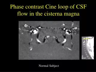
CSF flows the aquaduct of Sylvuis to the cisterna magna and then to the cisterna basalis
CSF flows from these basal cisterns past the tentorial notch and over the cerebral hemispheres
CSF passes through the arachnoid villi and into the superior saigittal sinus Superior sagittal sinus drains into the Venous Confluence which leads back to the circulatory system
The movement should not be thought of as a slow and steady flow, but rather as a constant ebb and flow due to arterial pulsations with a small net movement. CSF makes its way from the basal cisterns into the subarachnoid space around the spinal cord, descending caudally to the lumbar cistern and ascending rostrally, where most of the fluid is returned to the venous system through the arachnoid villi that penetrate the dural sleeves. Additional notes
The fate of 3H-water in CSF Space • Distribution of CSF ascribed to three processes (Riggs 1972) • Bulk Flow: fluid flows unidirectional, carrying all dissolved substances with the same speed • Mixing: Pressure waves disperses substances in all directions • Diffusion: Only efficient across very short distances • Since CSF is made up of over 99% CSF, labeled water used to investigate CSF flow (Bulat 1993)
Destination of 3H-water after 3 hrs Concentration in confluence of sinuses 5x higher than in cisterna magna
Bulat surmises that this indicates rapid passage of 3H-water from arterial blood into CNS and CSF – that this rapid passage can only be attributed to microvessel transportation. After three hours it is quite possible that CSF has accumulated in the confluence of sinuses via the cistern system – although Bulat showed a marked difference after one hour this is not a small enough discretization to by physiologically relevant. 3H-water lateral ventricle infusioninterpretation
The fate of 3H-inulin in CSF Space • A macromolecule (m.w. 5,500) which passes poorly across CNS capillaries (Crone, 1963) • Over time, 3H-inulin injected into money CSF becomes equal to that in the parenchyma – the cells that surround neural blood vessels (Kimelberg et. Al., 1978)
3H-inulin injection into CM collects in LSS Where is the inulin going?
Bulat’s Conclusion vs my own on 3H-inulin fate • Bulat believes the relatively straight spinal subarachnoid space, where systolic-diastolic mixing is prominent, explains how 3H-inulin can move so quickly - 70cm from CM to LSS. • Constricted radius may also contribute to high velocity, as well as increased absorption into LSS parenchyma, is there a higher concentration of subarachnoid villi in the LSS than the CSS?
Aquaduct of Sylvius occlusion in cats • 120 minutes after occlusion was introduced, cat brain exhibited no transmantle pressure, i.e. no difference in pressure between the lateral ventricles and the cisterna magna. • Lack of transmantle pressure observed in cats without the occlusion as well. • Bulat concludes that CSF does not flow through the Sylvian canal, that CSF is transported primarily by periventriculr capillaries.
Bulat’s theories are based on circumstantial evidence, no direct data Need quantitative data to support claim that subarachnoid granulations have ‘minimal surface area’ Need better modeling to predict pulsations and velocity of CSF – is one hour appropriate? Need more data on CSF velocities and distances Some current brain physiology models note the presence of a lateral aperture leading from the fourth ventricle to the pontine cistern – possible explanation for Sylvius occlusion experiment Measurements of transmantle pressures when periventricular capillaries or parenchyma are blocked from transporting CSF Conclusions