MYOPIA
MYOPIA. DR HASNAIN UL HAQ EYE SURGEON. DEFINITION.

MYOPIA
E N D
Presentation Transcript
MYOPIA DR HASNAIN UL HAQ EYE SURGEON
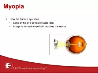
DEFINITION • Nearsightedness, or myopia, as it is medically termed, is a vision condition in which close objects are seen clearly, but objects farther away appear blurred. Nearsightedness occurs if the eyeball is too long or the cornea, the clear front cover of the eye, has too much curvature. As a result, the light entering the eye isn’t focused correctly and distant objects look blurred.
CAUSES • Nearsightedness is a very common vision condition affecting nearly 30 percent of the pak population. Some research supports the theory that nearsightedness is hereditary. There is also growing evidence that it is influenced by the visual stress of too much close work.
CAUSE • The exact cause of nearsightedness is unknown, but two factors may be primarily responsible for its development: • heredity • visual stress • There is significant evidence that many people inherit nearsightedness, or at least the tendency to develop nearsightedness. If one or both parents are nearsighted, there is an increased chance their children will be nearsighted. • Even though the tendency to develop nearsightedness may be inherited, its actual development may be affected by how a person uses his or her eyes. Individuals who spend considerable time reading, working at a computer, or doing other intense close visual work may be more likely to develop nearsightedness.
CONTINUE • Generally, nearsightedness first occurs in school-age children. Because the eye continues to grow during childhood, it typically progresses until about age 20. However, nearsightedness may also develop in adults due to visual stress or health conditions such as diabetes.
causes • There are three ways for an eye to become myopic: • The front surface of the eye (the cornea) is too curved and, therefore, too powerful. • The eyeball itself is too long. • A combination of both of the above.
Optics of myopia • The basic cause of myopia is increase in size of eyeball so that the light rays come to focus on the front of cornea.
Types • Simple • Pathological or degenerative or high mopia
SIMPLE MYOPIA • MOST COMMON TYPE THE EYEBALL IS INCREASED SLIGHTLY 1MM INCREASE IN EYEBALL SIZE LEADS TO 3D MYOPIA
Pathological myopia • In the more severe chronic cases ("degenerative" or "pathological" myopia), there is the possibility of sight loss. The deformation of the eye creates stress on the retina, which can become damaged or detached, and this can then provoke additional changes. This is especially true in degenerative myopia, which can lead to macula problems (not to be confused with age-related macular degeneration
continue • Degenerative myopia is more severe than other forms of myopia and is associated with retina changes, potentially causing severe vision loss. It progresses rapidly, and visual outcome depends largely on the extent of fundus and lenticular changes. The diagnosis of degenerative myopia is accompanied by characteristic chorioretinal degenerations. Pathologic myopes, particularly those with higher refractive errors, are at risk for retinal detachment and macular changes.
pathology • Vitreous liquefaction and posterior vitreous detachment • Peripapillary atrophy appearing as temporal choroidal or scleral crescents or rings around the optic disc • Lattice degeneration in the peripheral retina • Tilting or malinsertion of the optic disc, usually associated with myopic conus • Thinning of the retinal pigment epithelium with resulting atrophic appearance of the fundus • Ectasia of the sclera posteriorly (posterior staphyloma) • Breaks in Bruch's membrane and choriocapillaris, resulting in lines across the fundus called "lacquer cracks" • Fuch's spot in the macular area.
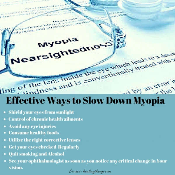
DIAGNOSIS • common sign of nearsightedness is difficulty with the clarity of distant objects like a movie or TV screen or the chalkboard in school. A comprehensive optometric examination will include testing for nearsightedness. An optometrist can prescribe eyeglasses or contact lenses that correct nearsightedness by bending the visual images that enter the eyes, focusing the images correctly at the back of the eye. Depending on the amount of nearsightedness, you may only need to wear glasses or contact lenses for certain activities, like watching a movie or driving a car. Or, if you are very nearsighted, they may need to be worn all the time.
VISUAL ACUITY • As part of the testing, letters on a distance chart are identified. This test measures visual acuity, which is written as a fraction such as 20/40. The top number of the fraction is the standard distance at which testing is performed, twenty feet. The bottom number is the smallest letter size read. A person with 20/40 visual acuity would have to get within 20 feet to identify a letter that could be seen clearly at forty feet in a “normal” eye. Normal distance visual acuity is 20/20, although many people have 20/15 (better) vision.
RETINOSCOPY • Using an instrument called a phoropter, an optometrist places a series of lenses in front of your eyes and measures how they focus light using a hand held lighted instrument called a retinoscope. The doctor may choose to use an automated instrument that automatically evaluates the focusing power of the eye. The power is then refined by patient’s responses to determine the lenses that allow the clearest vision.
TREATMENT • eyeglasses • contact lenses • laser and other refractive surgery procedures • vision therapy for persons with stress-related nearsightedness.
EYEGLASSES • Eyeglasses are the primary choice of correction for persons with nearsightedness. Generally, a single vision lens is prescribed to provide clear vision at all distances. However, for patients over about age 40, or children and adults whose nearsightedness is due to the stress of near vision work, a bifocal or progressive addition lens may be needed. These multifocal lenses provide different powers or strengths throughout the lens to allow for clear vision in the distance and also clear vision up close. • Eyeglasses are frequently used to correct myopia. • A large selection of lens types and frame designs are now available for patients of all ages. Eye glasses are no longer just a medical device that provides needed vision correction, but can also be a fashion statement. They are available in a wide variety of sizes, shapes, colors and materials that not only correct for vision problems but also may enhance appearance.
CONTECT LENSES • For some individuals, contact lenses can offer better vision than eyeglasses. They may provide clearer vision and a wider field of view. However, since contact lenses are worn directly on the eyes, they require regular cleaning and care to safeguard eye hea
PRK • Nearsightedness can also be corrected by reshaping the cornea using a laser beam of light. Two commonly used procedures are photorefractive keratectomy (PRK) and laser in situ keratomileusis (LASIK). • In PRK, a laser is used to remove a thin layer of tissue from the surface of the cornea in order to change its shape and refocus light entering the eye. There is a limit to how much tissue can safely be removed and therefore the amount of nearsightedness that can be corrected.
LASIK • LASIK does not remove tissue from the surface of the cornea, but from its inner layers. To do this, a section of the outer corneal surface is cut and folded back to expose the inner tissue. Then a laser is used to remove the precise amount of corneal tissue needed to reshape the eye, and then the flap of outer tissue is placed back in position to heal. The amount of nearsightedness that LASIK can correct is limited by the amount of corneal tissue that can be removed in a safe manner.