Download
1 / 15
150 likes | 396 Views
Stay updated on the latest treatment guidelines for mandibular third molars, including indications and risks, with a comparison of NICE and SIGN recommendations. Learn about potential complications, such as nerve injuries and dry socket, and the role of Cone Beam Computed Tomography in treatment planning.

E N D
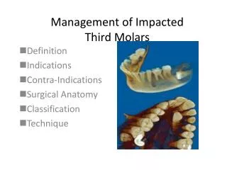
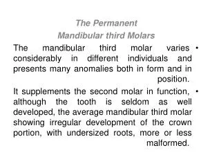
Treatment Planning for Mandibular Third Molars Catriona Jack
Current Guidance (NICE 2000) • Stressed that prophylactic surgical removal of impacted third molars should be discontinued on the NHS • Third molar surgery should be limited to patients with pathology • SIGN Guidance was withdrawn in 2015 to be revised • Since the implementation of NICE 2000 the incidence of distal caries in mandibular second molars has risen from 5% to 19%
Indications for Removal (NICE 2000) • Unrestorable caries in the third molar • Untreatable pulpal or periapical pathology • Cellulitis, abscess and osteomyelitis • Resorption of tooth or adjacent teeth • Diseases of the follicle including cysts or tumours • Teeth impeding surgery or in the field of tumour resection • Severe or multiple episodes of periodontitis
Differences between NICE and SIGN • SIGN included some indications for prophylactic removal: • Before radiotherapy or cardiac surgery • Periodontal disease of the second molar due to the position of the third molar • Patient occupation or lifestyle which prohibit access to regular dental care • Caries in the second molar which would be unrestorable without the removal of the third
Risks of Treatment • Post-op pain • Swelling • Trismus • Infection • Dry socket • Fractured Mandible Inferior alveolar nerve damage Lingual nerve damage
Post operative Infection: • Diagnosed by presence of suppuration, lymphadenopathy or systemic signs of infection • Cochrane review showed incidence of 10% in healthy patients, 25% in immunocompromised • Coulthard et al quote 3-4.7% • Mandibular Fracture: • A rare but serious complication (0.0049%) • Many occur 2-3 weeks post-op during mastication
Dry Socket • Severe pain following loss or lack of formation of blood clot in socket • Greatly varying quotes of incidence from 10% to 2% • Risk factors include: • Female • Difficulty of extraction (surgical extraction 9x greater risk than forceps extraction) • Expertise of surgeon • Smoking (3x greater risk) • Post-operative care
Temporary or permanent altered sensation to the areas supplied by the inferior alveolar or lingual nerves Altered sensation includes loss on sensation, tingling, pain or any abnormal sensation in the areas supplied by the inferior alveolar or lingual nerves Altered sensation lasting more than 6 months is usually categorised as permanent Nerve Injuries
Lingual Nerve • Temporary damage in 0-15% of cases • Permanent damage in 0-2% of cases • Large spread of figures due to varying techniques, lingual retraction a more historic technique where a retractor was used to protect the lingual nerve during sectioning, shown to increase the risk of temporary damage but reduce the risk of permanent damage
Inferior Alveolar Nerve • Temporary damage in 5% of cases • Permanent damage in 0.2% of cases • Incidence rises to when third molar is positioned in close proximity to the inferior alveolar nerve: • Temporary damage in 19% of cases • Permanent damage in 2% of cases • Radiographic signs of close relationship: • Interruption of white lines • Darkening of root • Deflection of root • Diversion of the inferior alveolar canal • Narrowing of the root
Cone Beam Computed Tomography • Allows 3D imaging of course of inferior alveolar nerve • Significant signs: • Narrowing of the canal • Direct contact of root and nerve • Lingual course of nerve • Intra-root position of the nerve Allows surgeon to treatment plan for extraction vs coronectomy One study showed that 12% of treatment plans were changed following CBCT
Dosages Effective dose of CBCT is 9.3-51.2μSv for small fields and 17.6-52.0μSv for full arches Effective dose for an OPT is up to 22μSv which is comparable An average risk of neoplasia formation of 2.7-9.8 per million
Coronectomy • Removal of crown only leaving roots which have not been mobilised • Sectioning of crown at 450 and removal of coronal portion so roots lie 3mm below alveolar bone and no enamel remains • Lingual nerve retraction described in some techniques, allows use of handpiece to section the crown, some force must be applied to roots if retraction is not used • Not indicated for mobile teeth or where caries extends into the pulp Risk of failed coronectomy varies between studies from 0% to 38%, likely due to difference in technique No significant difference in incidence of post-op pain and dry socket between coronectomy and surgical removal