Download

1 / 69
690 likes | 709 Views
Learn about bacterial cell walls, Gram staining, distinctive characteristics of Gram-negative bacteria, and enterobacteriaceae infections. Discover the significance of the oxidase and catalase tests in identifying bacteria. Dive into details about common Gram-negative infections like E.Coli, Salmonella, and Shigella.

E N D
Gram Negative Stains and Infections Presented by: Bryanna Gray Contact Information: 240-441-9227 Bryanna.Gray@yahoo.com
Bacterial Cell Wall Bacterial cells are covered by a cell envelope that is composed of a cell membrane and a cell wall. The cell membrane is a phospholipid bilayer that regulates the transport of molecules into and out of the cell. This is the weak structure that would burst from the osmotic pressure without reinforcement. 3 Primary Functions: • Provide Semi-Permeable Barrier Prevent loss of water Desirable substance passage • Protect against changes in osmotic pressure Main homeostasis and prevent bursting • Prevent Digestion by the host enzyme.
FUN FACT • The chemical composition of the bacterial cell wall is dramatically different from the mammalian cell wall and this provides a number of potentially attractive targets for the selective chemotherapy of the bacterial infections.
GRAM STAINING classifying BACTERIUM
Gram Staining Definition: a staining technique for the preliminary identification of bacteria, in which a violet dye(crystal violet) is applied, followed by a decolorizing agent(grams alcohol or ethanol/acetone) and then a red dye(safranin). The cell walls of certain bacteria (denoted Gram-positive ) retain the first dye and appear violet, while those that lose it (denoted Gram-negative ) appear red. Also called Gram's method. Inventor: Hans Christian Gram Purpose of Conducting a Gram Stain: Helps determine the identity of bacterial sample
4 Steps to Gram Staining Pour on crystal violet stain (a blue dye) and wait 60 seconds. Wash off with water and flood with iodine solution.Wait60 seconds. Wash off with water and then "decolorize" with 95% alcohol. Finally, counter-stain with safranin (a red dye). Wait 30 seconds and wash off with water.
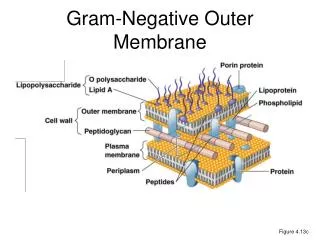
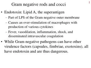
Gram Negative Stain Explanation • Crystal violet attaches to lipopolysaccharide layer Lipopolysaccharide: (LPS), also known as lipoglycans and endotoxins, are large molecules consisting of a lipid and a polysaccharide composed of O-antigen, outer core and inner core joined by a covalent bond they are found in the outer membrane of Gram-negative bacteria and elicit strong immune responses in animals. • Iodine acts as mordant • Grams alcohol washes away LPS layer & with it the stain • Safraninstains peptidoglycan Outermost layer lipopolysaccharide layer peptidoglycan layer Phospholipid bilayer cytoplasm
Review of Lab Test/Classifications Oxidase Test OXYGEN CLASSIFICATIONS catalase TEST
Oxidase Test The oxidase test is used to identify bacteria that produce cytochrome c oxidase, an enzyme of the bacterial electron transport chain. Bacteria that are oxidase positive are aerobic, and can use oxygen as a terminal electron acceptor in respiration.
Catalase Test The catalase test is used to differentiate staphylococci (catalase-positive) from streptococci (catalase-negative). The presence of catalase enzyme in the test isolate is detected using hydrogen peroxide. If the bacteria possess catalase(catalase-positive), when hydrogen peroxide is added bubbles of oxygen are observed.
Gram Negative Infections Enterobacteriaceae Genus
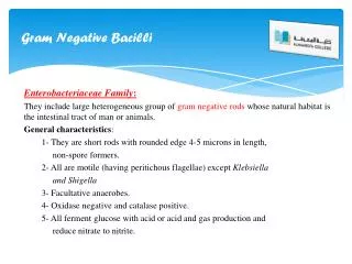
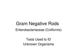
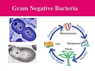
Characteristics of Enterobacteriacea Gram Negative Bacilli Facultative anaerobes Ubiquitous -Except for few, most are present in the intestinal tract of animals and humans as commensal flora; therefore, they are sometimes called “fecal coliforms” -Some live in water, soil and sewage
Biochemical Properties include: Oxidase Negative Catalase Positive Ferment glucose (dextrose) Reduces nitrates to nitrites
Entry of Enterobacteriaceae Contaminated medical devices Surgical site infections Intravenous (IV) Urinary Catheters Colonization of colon, perineum, urethra Pneumonias Contaminated food **Large numbers usually needed**
Evasion Capsule Anti-phagocytic Antigenic phase variation Altered expression of capsule flagella Plasmids Anti-microbial resistance
EnterobacteriaceaeInfections: E.Coli: Gastroenteritis Salmonella:a bacterium that occurs mainly in the intestine, especially a serotype causing food poisoning Shigella:a bacterium that is an intestinal pathogen of humans and other primates, some kinds of which cause dysentery
Escherichia coli • Most significant species in the genus • Natural inhabitant of the GI tract • Common isolate from colon flora • Ferments glucose(+) and lactose (+) • Positive indole and methyl red tests • Usually motile • Green Metallic Sheen on EMB agar
E.ColiExtraintestinal Infections Urinary Tract Infection Pulmonary infection Bacteremia Meningitis
E.Coli Intestinal Infections • Diarrhea • Hemolytic-uremic syndrome • Dysentery • Groups –based on how they cause illness • ETEC -Enterotoxigenic • EPEC -Enteropathogenic • EHEC -Enterohemorrhagic • EIEC -Enteroinvasive
EnterotoxigenicE.coli (ETEC) • Enterotoxigenic (ETEC) – “traveler’s diarrhea”; watery diarrhea without blood, nausea and vomiting; self-limiting; usually not identified, other than patient history and lactose-positive organisms cultured on differential media • Colonization Factor Antigens CFA/I,CFA/II -Facilitates bacteria adhesion • Entertoxins -Heat Labile -Heat Stable
Heat Labile In medicine, the term "labile" means susceptible to alteration or destruction. For example, a heat-labile protein is one that can be changed or destroyed at high temperatures. The opposite of labile in this context is "stable. Heat Labile A-B Toxin
Heat Stable Heat-stable enterotoxins (STs) are secretory peptides produced by some bacterial strains, such as enterotoxigenic Escherichia coli which are in general toxic to animals. These peptides keep their 3D structure and remain active at temperatures as high as 100 °C.
EnteropathogenicE.Coli (EPEC) • Strain of Escherichia coli in which organisms adhere to small bowel mucosa and produce characteristic changes in the microvilli. • This strain produces symptomatic, sometimes serious, gastrointestinal illnesses, especially severe in neonates and young children; • Responsible for Pediatric Diarrhea (symptoms: N/V, fever, watery diarrhea) • Entertoxins: -Heat Labile -Heat Stable
EPEC in small intestines Pathogenic Mechanism Attaching and effacing lesions Microbes bind to the epithelial cells via bundle-forming pilli (BFP) Loose, then tight binding Effacement lesion Associated pedestal formation
EnteroinvasiveE.Coli (EIEC) EIEC characteristics: • Dysentery with bowel penetration • Invasion and destruction of intestinal mucosa • Watery diarrhea with blood • Does NOT ferment lactose • Identified via DNA probes
EnteroinvasiveE.Coli (EIEC) Pathogenic Mechanism Invades intestinal epithelial cells Lyses the phagosomal vacuole Moves through the cytoplasm Spreads to adjacent cells
EnterohemorrhagicE.Coli (EHEC) • Enterohemorrhagic (EHEC serotype 0157:H7) • Associated with dysentery, hemorrhagic colitis, hemorrhagic diarrhea and hemolytic-uremic syndrome (HUS), which includes low platelet count, hemolytic anemia, and acute renal failure • Symptoms: Diarrhea and Bloody Diarrhea • Potentially fatal, especially in young children • Undercooked hamburger, unpasteurized milk and apple cider have spread the infection • Does NOT ferment sucrose • Identified by serotyping
EnterohemorrhagicE.Coli (EHEC) Pathogenic Mechanism Vero Toxin/ Shiga-like toxin AB Toxin-inhibits protein synthesis (similar to Shiga toxin) Acid Resistant Low infectious dose
Escherichia coli TREATMENT GI Infections Mainly Rehydration Therapy Antibiotics & Antidiarrheals are not recommended Most people recover within 5 - 10 days w/o medicine UTI and Systemic Infections- use antibiotics Sulfamethoxazole-Trimethoprim(Bactrim) Nitrofurantoin (Macrobid, Furadantin, Macrodantin)
Salmonella sp. Rod-shaped bacilli Lactose (-) H2S (+) Two Species of interest: S. typhi and S. typhosa May enter the digestive tract of humans/other mammals in contaminated food and cause abdominal pains and violent diarrhea Appears grayish on EMB agar.
Salmonella sp, Effects Diseases Gastroenteritis Septicemia Enteric Fever Carrier State Symptoms Fever Abdominal cramps Dysentery (blood diarrhea)
Salmonella sp. Pathogenic Effects Gastroenteritis Adhesion to microvilli Cellular invasion Survival in vesicle Multiplication ESCAPE Septicemia (all of the above +) Silent Septicemia Uptake via Macrophages Survival in Macrophages vesicle SYSTEMIC Enteric Fever (all of the above +) Muliplication in lymph nodes, spleen, liver macrophages Bacteremia release from macrophages to blood High Fever (Endotoxin) Reinvasion of intestine via gall bladder Carrier State Established gall bladder infection
Salmonella sp. Treatment Self-limiting Hydration Usually resolves in 5-7 days Systemic Infection Ampicillin Sulfamethoxazole-Trimethoprim (Bactrim) Ciprofloxacin
Shigella sp. Lactose (-) H2S (-) Non-motile Three Species of interest: S. dysenteriae, S. sonnei & S. fexneri Symptoms: Fever, abdominal cramps, dysentery. Presentation is similar to Salmonella…WHY?
Shigella Entry Fecal-oral transmission Acid Resistant- small numbers needed
Shigella Evasion Cellular invasion-escape from endocytic vesicle, multiplication in the cytoplasm Spread to adjacent cell
ShigellaTreatment Self-limiting Usually resolves within 5-7 days Hydration Ampicillin, Bactrim and Ciprofloxacin can be used
Gram Negative Infections Neisseria genus
Neisseria Gonorrhoeae Gram – diplococci Aerobic Cytochrome oxidase (+) Maltose (-) N.Gonorrhoeae: Gonorrhea oldest STI, venereal disease involving inflammatory discharge from the urethra or vagina
Entry Infects mucosal surfaces Ex. cervix, urethra, rectum, oropharynx, nasopharynx, conjunctiva
Evasion Capsule (antiphagocytic)
N. Gonorrhea Pathological Effects Gonorrhea Symptomatic or asymptomatic Transmission Sexual Contact Newborns
N. Gonorrhea Pathological Effects (continued) Men Women Asymptomatic infection Acute urethritis Purulent exudate w/ dysuria, polyuria, HA, fever Anterior to posterior urethral infection Glands, ducts, and vesicles may become infected Chronic infection Prostate, seminal vesicles, and epididymitis may become infected Asymptomatic carriers Acute gonorrhea infection (lower tract) Endocervix (traditional site of infection) Acute symptoms Ab/pelvic pain, vaginal discharge/dysuria Chronic Gonorrhea infection Tenderness in lower abdomen, inflammation of the urinary tract Pelvic Inflammatory Disease (PID)