Download
1 / 15
160 likes | 360 Views
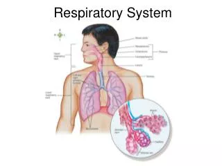
Respiratory System. Respiratory Tract. Conducting passageways carrying air to and from the alveoli Upper respiratory passages filter and humidify incoming air Nasal Cavity Larynx Pharynx Nasopharynx Oropharynx Laryngopharynx

E N D
Respiratory Tract • Conducting passageways carrying air to and from the alveoli • Upper respiratory passages filter and humidify incoming air • Nasal Cavity • Larynx • Pharynx • Nasopharynx • Oropharynx • Laryngopharynx • Lower passageways include delicate conduction passages and alveolar exchange surfaces • Trachea • Lungs • Bronchi • Bronchioles • Alveoli • Diaphragm Pathway nasal cavities (or oral cavity) → pharynx → trachea → primary bronchi (right & left) → secondary bronchi → tertiary bronchi → bronchioles → alveoli
Nasal Cavity/Nose • Functions • Provides an airway for respiration • Moistens and warms entering air • Filters and cleans inspired air • Resonating chamber for speech • Detects odors in the airstream • Selected anatomical features • Vibrissae(guard hairs) – stiff hairs that filter large particles from the air • Nasal cilia– hair-like projections that propel trapped particles towards the throat for digestion by digestive enzymes • Rich supply of capillaries warm the inspired air • Nasal conchae– folds in the mucous membrane that increase air turbulence and ensures that most air contacts the mucous membranes • Olfactory mucosa – mucous membranes that contain smell receptors • Respiratorymucosa – pseudostratified ciliated columnar epithelium containing goblet cells that secrete mucus • Mucus • Stickiness traps inhaled particles • Lysozyme kills bacteria • Lymphocytes and IgA antibodies protect against bacteria
Upper Respiratory • Three regions of the pharynx • Nasopharynx– air passage (pseudostratified columnar epithelium) • Oropharynx – passageway for air, food, and drink (stratified squamous epithelium) • Laryngopharynx – passageway for air, food, and drink (stratified squamous epithelium)
Larynx • Functions • 1. Keeps food and drink out of the airway • 2. Sound production • 3. Acts as a sphincter during abdominal straining (ex. during defecation and heavy lifting)
Larynx • Selected anatomical features • Nine c-rings of hyaline cartilage form the framework of the larynx • Muscular walls aid in voice production and the swallowing reflex • Glottis – the superior opening of the larynx • Epiglottis – prevents food and drink from entering airway when swallowing • False vocal cords – aid in closing the glottis when swallowing • True vocal cords – produce sound when air passes between them • The shorter and thinner these membranes are, the faster air moves over them – produces high pitched sounds • The longer and thicker these membranes are, the slower air moves over them – produces low pitched sounds
Trachea • Functions • Air passageway • 2. Cleans, warms, and moistens incoming air • 3. Selected anatomical features • Rings of hyaline cartilage – reinforce the trachea and keep it from collapsing when you inhale • Ciliated pseudostratified epithelium – traps inhaled debris and propels mucus up to the pharynx where it is swallowed
Bronchi • Function - Solely an air passageway • Selected anatomical features • 1. Left and right primary bronchi branch off from trachea • 2. Once the left and right primary bronchi enter the lungs they are subdivided into smaller tubes: • Secondary bronchi (one for each lobe) → tertiary bronchi → bronchioles → terminal bronchioles → respiratory bronchioles → alveolar ducts → alveolar sacs • 3. Alveolar sacs are clusters of alveoli • --Alveoli are the site of gas exchange • --Surfactant prevents from collapsing due to tension • --Other tissue types present in the alveoli • a. Smooth muscle rings aid in resistance to air flow • b. Elastic connective tissue fibers aid in expelling air from the lungs
Pulmonary arteriogram Lung bronchiogram


