Download
1 / 25
330 likes | 694 Views
P53, Apoptosis, Cancer, More Regulation . G1 checkpoint. Controlled by G1 Cdk-cyclin G1 cyclin levels also vary with the cell cycle Many additional levels of phosphorylation, dephosphor-ylation regulate. E2F. P. E2F. P. E2F. E2F. E2F. P. E2F. E2F. E2F. P. G1. Cyc D. cdk4. S.

E N D
G1 checkpoint • Controlled by G1 Cdk-cyclin • G1 cyclin levels also vary with the cell cycle • Many additional levels of phosphorylation, dephosphor-ylation regulate.
E2F P E2F P E2F E2F E2F P E2F E2F E2F P G1 Cyc D cdk4 S P pRB Cyc E cdk2
Growth Factor Signaling Through the Ras Pathway crossing of G1 checkpoint Ras*, Raf* MAPK cascade Activation of nuclear TFs Activation of G1 Cdk cyclin genes: G1 S
G2 Checkpoint Control by MPF • Active MPF = Mitotic Cdk + mitotic cyclin • Cdk is cyclin-dependant kinase • MPF controls G2 M by phosphorylating and activating proteins involving in: • Chromosome condensation • Nuclear envelope breakdown • Spindle assembly • It’s own self-destruction
Check Separase Check APC Separase Separase Separase point Securin Securin point Cohesin Cohesin prometaphase metaphase anaphase Sister chromatid separation from prometaphase to anaphase APC Ub Ub Ub Securin
Spindle Assembly Checkpoint Controls Metaphase Anaphase • MPF (+) anaphase promoting complex, which destroys: • Securin, which allows separin protease to cleave cohesin. • Mitotic cyclin, which causes loss of MPF activity, leading to chromosome decondensation and envelope reformation. (+)
Section 20.2: Control of Mitosis by Cyclins and MPF Activity Maturation promoting factor (MPF) is a protein kinase (requiring a mitotic cyclin) that stimulates mitosis, and hence is also called mitosis promoting factor. The increase and decrease in MPF activity during the cell cycle in the early embryo reflects the cyclic synthesis and degradation of mitotic cyclins. The anaphase-promoting complex/cyclosome (APC/C) is a ubiquitin ligase that recognizes the destruction box sequence in mitotic cyclins, marking them for degradation in late anaphase and thus terminating mitosis. Deactivation of APC/C (by G1-cyclin/CDK) in G1 permits mitotic (and S phase) cyclins to accumulate, allowing the cell cycle to continue.
P53 DEF: A gene which encodes a protein that regulates all growth and is able to cause potential cancerous cells to destroy themselves. The gene is an antioncogen. F(x): Includes regulation of critical cellular function involving the G1 and G2 cell-cycle check points in response to DNA damage and apoptosis induced by certain stimuli, such as DNA damaging agents and hypoxia. Additional F(x): The tumor suppressor gene functions as the “guardian of the genome” plays a pivotal roe in “sending” damaged DNA and in making critical decisions of whether a cell should repair the damaged DNA.
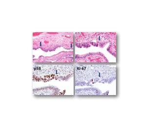
The tumor suppressor gene p53 is located at chromosomes region 17p13 and is one of the most frequently mutated gene in human cancers.
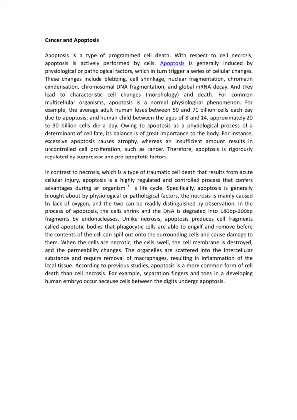
APOPTOSIS Programmed cell death Orderly cellular self destruction Process: as crucial for survival of multi-cellular organisms as cell division
IMPORTANCE OF APOPTOSIS • Important in normal physiology / development • Development: Immune systems maturation, Morphogenesis, Neural development • Adult: Immune privilege, DNA Damage and wound repair. • Excess apoptosis • Neurodegenerative diseases • Deficient apoptosis • Cancer • Autoimmunity
STAGES OF CLASSIC APOPTOSIS Healthy cell DEATH SIGNAL (extrinsic or intrinsic) Commitment to die (reversible) EXECUTION (irreversible) Dead cell (condensed, crosslinked) ENGULFMENT (macrophages, neighboring cells) DEGRADATION
APOPTOSIS: Role in Disease TOO MUCH: Tissue atrophy Neurodegeneration Thin skin etc TOO LITTLE: Hyperplasia Cancer Athersclerosis etc
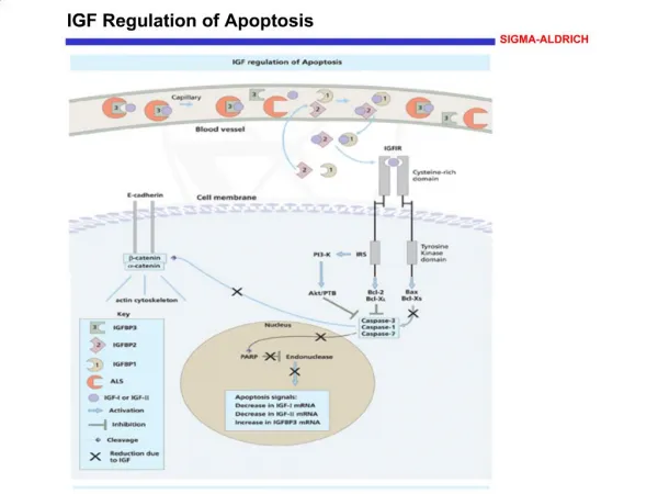
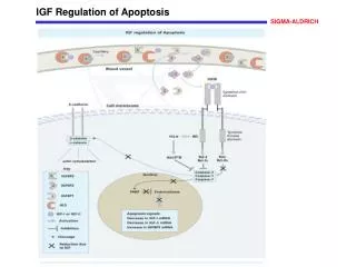
APOPTOSIS: Role in Disease: Cancer Apoptosis eliminates damaged cells (damage => mutations => cancer Tumor suppressor p53 controls senescence and apoptosis responses to damage Most cancer cells are defective in apoptotic response (damaged, mutant cells survive) High levels of anti-apoptotic proteins or Low levels of pro-apoptotic proteins ===> CANCER
OPTIMAL FUNCTION (HEALTH) APOPTOSIS AGING APOPTOSIS Neurodegeneration, cancer, …..
P53 & Apoptosis p53 first arrests cell growth between G1 S This allows for DNA repair during delay If the damage is too extensive then p53 induces gene activation leading to apoptosis (programmed cell death)
Cancer: Benign • Benign: localized and of small size • Cells that closely resemble, and may function, like normal cells • Become problems due to sheer bulk or due to secretions (e.g. hormones)
Cancer : Malignant Malignant tumors: high rate of division, properties may vary compared to cells of origin. Most malignant cells become metastatic Invade surrounding tissue and establishment of secondary areas of growth: Metastasis