Elbow Joint
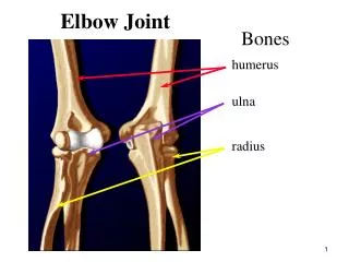
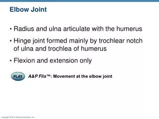
Elbow Joint. Radius and ulna articulate with the humerus Hinge joint formed mainly by trochlear notch of ulna and trochlea of humerus Flexion and extension only. PLAY. A&P Flix ™: Movement at the elbow joint. Articular capsule. Synovial membrane. Humerus. Synovial cavity.

Elbow Joint
E N D
Presentation Transcript
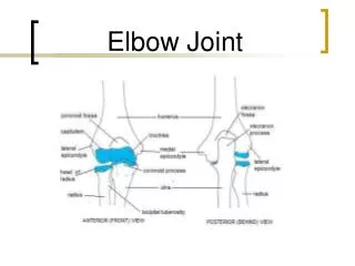
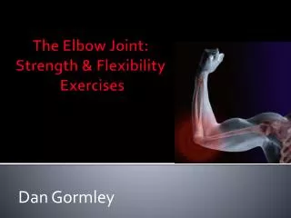
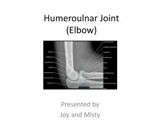
Elbow Joint • Radius and ulna articulate with the humerus • Hinge joint formed mainly by trochlear notch of ulna and trochlea of humerus • Flexion and extension only PLAY A&P Flix™: Movement at the elbow joint
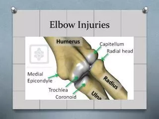
Articular capsule Synovial membrane Humerus Synovial cavity Articular cartilage Fat pad Coronoid process Tendon of triceps muscle Tendon of brachialis muscle Ulna Bursa Trochlea Articular cartilage of the trochlear notch (a) Median sagittal section through right elbow (lateral view) Figure 8.11a
Elbow Joint • Anular ligament—surrounds head of radius • Two capsular ligaments restrict side-to-side movement: • Ulnar collateral ligament • Radial collateral ligament
Humerus Anular ligament Radius Lateral epicondyle Articular capsule Radial collateral ligament Olecranon process Ulna (b) Lateral view of right elbow joint Figure 8.11b
Articular capsule Anular ligament Humerus Coronoid process Medial epicondyle Ulnar collateral ligament Radius Ulna (d) Medial view of right elbow PLAY Animation: Rotatable elbow Figure 8.11d
Hip (Coxal) Joint • Ball-and-socket joint • Head of the femur articulates with the acetabulum • Good range of motion, but limited by the deep socket • Acetabular labrum—enhances depth of socket PLAY A&P Flix™: Movement at the hip joint: An overview
Coxal (hip) bone Articular cartilage Ligament of the head of the femur (ligamentum teres) Acetabular labrum Femur Synovial cavity Articular capsule (a) Frontal section through the right hip joint Figure 8.12a
Hip Joint Reinforcing ligaments: • Iliofemoral ligament • Pubofemoral ligament • Ischiofemoral ligament • Ligamentum teres
Iliofemoral ligament Ischium Ischiofemoral ligament Greater trochanter of femur (c) Posterior view of right hip joint, capsule in place PLAY Animation: Rotatable hip Figure 8.12c
Iliofemoral ligament Anterior inferior iliac spine Pubofemoral ligament Greater trochanter (d) Anterior view of right hip joint, capsule in place Figure 8.12d
Temporomandibular Joint (TMJ) • Mandibular condyle articulates with the temporal bone • Two types of movement • Hinge—depression and elevation of mandible • Gliding—e.g. side-to-side (lateral excursion) grinding of teeth • Most easily dislocated joint in the body
Mandibular fossa Articular tubercle Zygomatic process Infratemporal fossa External acoustic meatus Lateral ligament Articular capsule Ramus of mandible (a) Location of the joint in the skull Figure 8.13a
Articular disc Articular tubercle Mandibular fossa Superior joint cavity Articular capsule Synovial membranes Mandibular condyle Ramus of mandible Inferior joint cavity (b) Enlargement of a sagittal section through the joint Figure 8.13b
Superior view Outline of the mandibular fossa Lateral excursion: lateral (side-to-side) movements of the mandible Figure 8.13c
Common Joint Injuries • Sprains • The ligaments are stretched or torn • Partial tears slowly repair themselves • Complete ruptures require prompt surgical repair • Cartilage tears • Due to compression and shear stress • Fragments may cause joint to lock or bind • Cartilage rarely repairs itself • Repaired with arthroscopic surgery
Torn meniscus Figure 8.14
Common Joint Injuries • Dislocations (luxations) • Occur when bones are forced out of alignment • Accompanied by sprains, inflammation, and joint immobilization • Caused by serious falls or playing sports • Subluxation—partial dislocation of a joint
Inflammatory and Degenerative Conditions • Bursitis • An inflammation of a bursa, usually caused by a blow or friction • Treated with rest and ice and, if severe, anti-inflammatory drugs • Tendonitis • Inflammation of tendon sheaths typically caused by overuse • Symptoms and treatment similar to bursitis
Arthritis • >100 different types of inflammatory or degenerative diseases that damage joints • Most widespread crippling disease in the U.S. • Symptoms; pain, stiffness, and swelling of a joint • Acute forms: caused by bacteria, treated with antibiotics • Chronic forms: osteoarthritis, rheumatoid arthritis, and gouty arthritis
Osteoarthritis (OA) • Common, irreversible, degenerative (“wear-and-tear”) arthritis • 85% of all Americans develop OA, more women than men • Probably related to the normal aging process
Osteoarthritis (OA) • More cartilage is destroyed than replaced in badly aligned or overworked joints • Exposed bone ends thicken, enlarge, form bone spurs, and restrict movement • Treatment: moderate activity, mild pain relievers, capsaicin creams, glucosamine and chondroitin sulfate
Rheumatoid Arthritis (RA) • Chronic, inflammatory, autoimmune disease of unknown cause • Usually arises between age 40 and 50, but may occur at any age; affects 3 times as many women as men • Signs and symptoms include joint pain and swelling (usually bilateral), anemia, osteoporosis, muscle weakness, and cardiovascular problems
Rheumatoid Arthritis • RA begins with synovitis of the affected joint • Inflammatory blood cells migrate to the joint, release inflammatory chemicals • Inflamed synovial membrane thickens into a pannus • Pannus erodes cartilage, scar tissue forms, articulating bone ends connect (ankylosis)
Rheumatoid Arthritis: Treatment • Conservative therapy: aspirin, long-term use of antibiotics, and physical therapy • Progressive treatment: anti-inflammatory drugs or immunosuppressants • New biological response modifier drugs neutralize inflammatory chemicals
Gouty Arthritis • Deposition of uric acid crystals in joints and soft tissues, followed by inflammation • More common in men • Typically affects the joint at the base of the great toe • In untreated gouty arthritis, the bone ends fuse and immobilize the joint • Treatment: drugs, plenty of water, avoidance of alcohol
Lyme Disease • Caused by bacteria transmitted by the bites of ticks • Symptoms: skin rash, flu-like symptoms, and foggy thinking • May lead to joint pain and arthritis • Treatment: antibiotics
Developmental Aspects of Joints • By embryonic week 8, synovial joints resemble adult joints • A joint’s size, shape, and flexibility are modified by use • Advancing years take their toll on joints: • Ligaments and tendons shorten and weaken • Intervertebral discs become more likely to herniate • Most people in their 70s have some degree of OA • Exercise that coaxes joints through their full range of motion is key to postponing joint problems