Download
1 / 25
260 likes | 501 Views
Pathology of the Gastrointestinal Tract Part 1 Small and Large Intestines. Grace Guzman, M.D. graceguz@uic.edu The Department of Pathology University of Illinois at Chicago. Atresia and stenosis. Congenital intestinal obstruction -Complete: Atresia -Incomplete: Stenosis

E N D
Pathology of the Gastrointestinal Tract Part 1Small and Large Intestines Grace Guzman, M.D. graceguz@uic.edu The Department of Pathology University of Illinois at Chicago
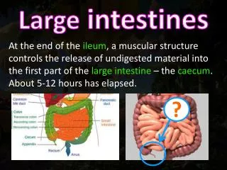
Atresia and stenosis • Congenital intestinal obstruction -Complete: Atresia -Incomplete: Stenosis • Duodenal: most common -Jejunum and ileum: equal -Rectum: rare • Developmental failure • intrauterine vascular accidents, or intussuception • Imperforate anus
Meckel Diverticulum • Persistence of omphalomesenteric duct (vitelline duct) • Disease of 2’s -2% of population (mostly asymptomatic) -M:F 2:1 -2” in length -2 ft of ileocecal valve -2 types of ectopic tissue in 1/2 of cases (gastric and pancreatic) -2 major complications (pain with inflammation; hemorrhage with ulcer)
Absence of ganglia -submucosal (Meissner) -myenteric (Auerbach) Alternating obstruction and diarrhea Aganglionic segment causes functional obstruction with distention proximal to aganglionic segment M:F 4:1 Down syndrome (10%)and (5%) serious neurologic abnormalities 1 in 5000 to 8000 Presents in neonatal period (failure to pass meconium; abdominal distention) Risk of perforation, sepsis, enterocolitis, fluid disturbances Acquired (Chagas disease) Congenital Aganglionic Megacolon“Hirschsprung Disease” Intestinal neuronal plexus develop from neural crest cells migrate to the bowel during development Sporadic Familial Gentic defects: Endothelin 3 GCDGF Receptor tyrosine kinase
Enterocolitis • Infectious • Necrotizing • Pseudomembranous Infectious -Viral (Rotavirus, Norwalk) -Bacterial E. coli; Shigella; V. Cholerae; C. difficile -Parasites and protozoa (nematodes; flatworms; protozoa -Giardia lambdia; E. histolytica)
Acute, necrotizing inflammation of small and/or large intestines Most common acquired GI emergency in premature or low birth weight neonate Mild GI symptoms or fulminant illness Multifactorial - immaturity of the gut’s immune system Release of cytokines and endotoxins damages mucosa and blood supply Terminal ileum or ascending colon Edema to necrosis to gangrenous bowel Necrotizing enterocolitis
Pseudomembranous colitis (antibiotic associated) Dx: C. difficile cytotoxin in stool Response to tx is usually prompt Relapse occurs in up to 25% of px • Yellow green false membrane (mixture of mucous and neutrophils) • Toxin produced by Clostridium difficile (acquired nasocomially in 20% of pxs in long term hospitalization) • Antibiotics allow overgrowth of C. difficile • Sudden onset of fever and diarrhea in a patient who is seriously ill or post operative who is receiving antibiotics • diarrhea, dehydration, shock death Exotoxin A and B binds to enteric receptors inactivates RhO cytoplasmic proteins causing injury to actin filaments and cell retraction
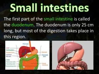
Malabsorption • Defect in the assimilation of food (digestion and absorption) • Intraluminal stage a. Secretory Phase (Chronic pancreatitis/insufficiency) b. Biliary Phase (Biliary obstruction due to calculus of or tumor) • Intestinal Stage (terminal digestion) a. Surface Phase (Celiac disease; bowel resection) b. Cellular Phase (Disaccharidase deficiency) • Removal Stage (transepithelial transport) a. Delivery Phase (Whipple diease)
Celiac sprue • Gluten, gliadin protein in wheat, oat, barley, and rye • hypersensitivity (immunologic) reaction to gluten • 90-95% - HLA DQ heterodimer in Ch 6 • Whites - rare in native Africans, Japanese, Chinese • Gluten - malabsorption -gluten free - improvement • Long term risk of malignancy -lymphoma (2X normal) • Distinct from Tropical sprue Celiac disease: loss of villi increased crypts, inflammation, intraepithelial lymphocytes, loss of brush border, goblet cells
Whipple disease • Rare • Gram positive rod shaped actinomycete: Tropheryma whippleli • Engulfed by macrophages (PAS positive diastase resistant) • Electron microscopy • M:F 10:1
Inflammation • 1. Miscellaneous -graft vs. host -drug induced -radiation enterocolitis -neutropenic colitis -diversion colitis • 2. Acute appendicitis -etiology: bacteria -fecalith impairing circulation, causing ischemia, necrosis and bacterial contamination -acute abdomen -RLQ pain-McBurney’s point -fever and leukocytosis
Inflammation3. Collagenous and lymphocytic colitis • Etiology: unknown • possibly auto-immune • chronic watery diarrhea in middle aged and older women • spectrum of disease ranging from increased intraepithelial lymphocytes to the presence of collagen band under the surface epithelium
Inflammatory bowel disease (IBD) - single term to collectively refer to either Crohn disease or ulcerative colitis Etiology unknown a. Genetic predisposition:HLA Class II locus on Ch 6 b. Abnormal host immunoreactivity Idiopathic Inflammatory Bowel disease
Crohn disease: Regional enteritis 1. Chronic inflammation involving all layers (transmural) of the SI • may occur at any point along the GI tract • primarily involving SI and LI 2. Mucosa shows linear ulceration and fistula 3. Segmental involvement/sparing • Serosal creeping fat
Crohn disease: Regional enteritis • Inflammation spread through the bowel wall to adjacent mesenteric fat • -characteristic non-caseating granulomas • tends to occur in young adults • increased incidence of cancer of SI and colon • diarrhea, crampy abdominal pain, fever • complications: fistula, obstruction, occult blood loss, Fe++ def anemia • malabsorption, malnutrition, weight loss
Ulcerative colitis 1. Inflammation primarily involving the mucosa of the colon 2. Diffuse, continuous inflammation that begins in the rectum and progresses proximally 3. Pseudopolyp formation 4. Bloody diarrhea, from ruptured vessels in inflamed mucosa • Toxic megacolon - rare complication - prominent dilatation and septic shock
Ulcerative colitis • Early phase: neutrophils accumulate within the depths of the crypts of Leiberkuhn forming crypt abscesses • Later phase: mucosa ulcerates and pseudo-polyps form • Late phase: after many years, mucosa becomes dysplastic, increasing risk of colon carcinoma
Transmural inflammation pseudopolyp granuloma diffuse skip lesions toxic megacolon creeping fat Primary Sclerosing Cholangitis fissures and fistulas Cancer at any point in GI tract Rectum Crohn UC Crohn UC Crohn UC Crohn both but more in UC Crohn both but more in UC Crohn UC Between Crohn and UC, this finding is more commonly seen in:
Vascular diseases:a. Ischemic bowel diseaseb. Angiodysplasiac. Hemorrhoids • Ischemic bowel disease • -blood clot in mesenteric artery causing ischemia, transmural infarction, necrosis of bowel, peritonitis a.embolus: superior mesenteric artery -source: embolus of heart (mural thrombus, valvular vegetation) b. thrombus (arterial; venous: ATIII def, cirrhosis, OC) c. hypoperfusion (non-occlusive): shock, CHF 50-75% death rate older px with cardiac, vasc disease D/Dx: IBD
Vascular diseases:a. Ischemic bowel diseaseb. Angiodysplasiac. Hemorrhoids • Angiodysplasia • -ectasia of veins • -prone to rupture • -GI bleeding • -Osler-Weber-Rendu syndrome (hereditary hemorrhagic telangiectasia) • Hemorrhoids • -dilated veins of hemorrhoidal plexus • -Internal • -External • -(BRBPR or streaks on stool), thrombosis, pain Prevalence:<1% 20% of significant LGI bleed 5% of population elevated venous pressure constipation straining venous stasis of pregnancy collateral channels in portal HTN rare under 30 except in pregnant women
Non-neoplastic bowel diseasesa. Diverticular diseaseb. Herniasc. Adhesionsd. Intussusceptione. Volvulus • Diverticular disease: • Diverticulosis and Diverticulitis • Acquired herniation • Most common in left colon; particularly sigmoid colon • Acute or chronic inflammation may occur • Perforation, peritonitis, fistula Acquired rare under 30 western pop over 60 prevalence: 50%
Non-neoplastic bowel diseasesa. Diverticular diseaseb. Herniasc. Adhesionsd. Intussuceptione. Volvulus • Hernias -Serosal lined out-pouching of peritoneum -Loop of intestines becomes trapped (incarcerated) within the hernia sac -Bowel compressed, twisted at the mouth of hernia, compromising blood supply - infarction (strangulation)
Non-neoplastic bowel diseasesa. Diverticular diseaseb. Herniasc. Adhesionsd. Intussuceptione. Volvulus • Adhesions • -string-like or band-like portions of scar tissue that form during healing after surgery or peritonitis • -may result in obstruction (kinking, compression)
Non-neoplastic bowel diseasesa. Diverticular diseaseb. Herniasc. Adhesionsd. Intussusceptione. Volvulus • Intussusception • -caused by an in-folding or telescoping of one segment of bowel into the adjacent distal segment • Infants and children: spontaneous and reversible • Adults: tumor is usually a lead point
Non-neoplastic bowel diseasesa. Diverticular diseaseb. Herniasc. Adhesionsd. Intussuceptione. Volvulus • Volvulus • -obstruction due to rotation or twisting of a loop of bowel around its mesenteric base of attachment • Sigmoid - most common site (cecum next)