Understanding Meiosis: The Process of Gamete Formation and Genetic Variation
Meiosis is a crucial cell division process that produces genetically unique sex cells known as gametes (sperm and eggs). This process reduces the chromosome number by half, resulting in haploid cells (23 chromosomes in humans), contrasting diploid body cells (46 chromosomes). Meiosis consists of two division rounds (Meiosis I and II), involving steps such as homologous chromosome pairing and crossing over for genetic variation. Sources of genetic diversity include crossing over, independent assortment, and random fertilization, leading to vast combinations of gametes.

Understanding Meiosis: The Process of Gamete Formation and Genetic Variation
E N D
Presentation Transcript
Cell Division Notes part 3: Meiosis
Meiosis • Meiosis is a type of cell division that is used to produce genetically unique gametes, or sex cells • Male gametes are sperm cells; female gametes are eggs • Gametes have only half the original number of chromosomes; this is known as being haploid (1n) versus the diploid (2n) body cells • n = number of chromosomes • Example: Human diploid number is 46; human haploid number is 23
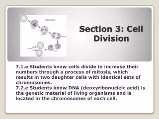
Meiosis results in 4 cells with half of the original chromosomes as the parent cell
2 rounds of division • Meiosis requires 2 rounds of division (MI and MII) • Homologous chromosomes are separated in the first round of division • Homologous chromosomes are matching pairs • Sister chromatids are separated in the second round of division Meiosis I Meiosis II
Steps of Meiosis I • STEP 1: PROPHASE I • Chromatin condenses; chromosomes become visible. Nuclear membrane breaks down; centrioles move apart. • Homologous pairs come together—form tetrads. • Crossing over occurs—homologous chromosomes “swap” genetic material
Steps of Meiosis I • STEP 2: METAPHASE I • Centrioles are at opposite ends of cell. • Spindle fibers attach to sister chromatids at kinetochore of the centromere. • Homologous chromosomes pair up at center of cell.
Steps of Meiosis I • STEP 3: ANAPHASE I • Homologous pairs are pulled apart, move to opposite ends of the cell by spindle fibers. (they are still in an “X” shape)
Steps of Meiosis I • STEP 4: TELOPHASE I/ CYTOKINESIS I (aka – INTERKINESIS) • Cell pinches into 2 cells
Steps of Meiosis II • STEP 5: PROPHASE II • 2 new cells, each with half the original number of chromosomes (haploid) and sister chromatids still attached. • Centrioles double and move apart; spindle fibers form again. • No nuclear membrane.
Steps of Meiosis II • STEP 6: METAPHASE IICentrioles are at opposite ends of cell. • Spindle fibers attach to sister chromatids at kinetochores of the centromere. • Sister chromatids line up at center of cell.
Steps of Meiosis II • STEP 7: ANAPHASE II • Sister chromatids are pulled apart, move to opposite ends of cell by spindle fibers.
Steps of Meiosis II • STEP 8: TELOPHASE II/CYTOKINESIS II • Nuclear membrane forms around chromosomes at each end of cell. • Each of the 2 cells pinch into 2 cells, making 4 haploid gametes.
Meiosis Animation • Stages of Meiosis • Images of Nondisjunction in Meiosis • When chromosomes do not separate as they should
Genetic Variation • 3 sources of genetic variation: many possible combinations of genes in sexual reproduction 1. Crossing over: swapping of genes in Prophase I of Meiosis; homologous chromosomes trade chromosome parts
Genetic Variation 2. Law of independent assortment: homologous chromosomes line up randomly and randomly separate to each new cell (some of mom’s chromosomes, some of dad’s) Animation
Genetic Variation 3. Random fertilization: many sperm could fertilize each egg, but only one will—the possibilities for fertilization are almost endless A zygote is formed by the random union of independently-produced gametes. For humans, the number of different gametes is 223 * 223, or 8,388,6082, giving 70,368,744,177,664 (70 trillion) possible combinations!
Sperm vs. egg cells • Sperm cells are much smaller than egg cells, and have a tail-like cell part called a flagellum. • In humans, both sperm and egg cells begin to develop by meiosis at puberty. • Sperm form in the testes; eggs form in the ovaries.