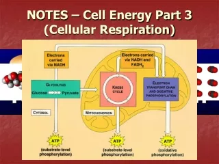
Brain Notes Part 3
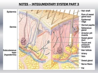
Brain Notes Part 3. The Cerebrum, Higher Brain Functions, and Brain Mapping. Surface of the Cerebral Hemispheres: Major anatomical landmarks on the surface of the left cerebral hemisphere are shown. The colored areas represent various motor , sensory, and association areas.

Brain Notes Part 3
E N D
Presentation Transcript
Brain Notes Part 3 The Cerebrum, Higher Brain Functions, and Brain Mapping
Surface of the Cerebral Hemispheres: Major anatomical landmarks on the surface of the left cerebral hemisphere are shown. The colored areas represent various motor, sensory, and association areas. Association areas = adjacent regions that interpret incoming data or coordinate a motor response. *Processing centers = “Higher-order” integrative centers that receive information from many different association areas; control extremely complex motor activities and perform complicated analytical functions; many of these centers are LATERALIZED (speech, writing, math comp, spatial understanding)
*Primary motor cortex (precentralgyrus, frontal lobe) – neurons direct voluntary movements by controlling somatic motor neurons in the brain stem and spinal cord. Functional areas of the Cerebrum
Functional areas of the Cerebrum *Somatic sensory association area (parietal lobe) – monitors activity in the primary sensory cortex; this area allows you to recognize a touch as light as the arrival of a mosquito on your arm. *Primary sensory cortex (postcentralgyrus, parietal lobe) – neurons in this region receive somatic sensory information from touch, pressure, pain, and temperature receptors; we are consciously aware of these sensations because the brain stem nuclei relay sensory information to the primary sensory cortex.
Functional areas of the Cerebrum *visual association area (occipital lobe) – is responsible for determining meaning of visual input.
Functional areas of the Cerebrum *General Interpretive Area [aka. Wernicke’s area] – receives information from all the sensory association areas; plays an essential role in your personality by integrating sensory info and coordinating access to complex visual and auditory memories; represented in LEFT hemisphere; (you can recognize the meaning of the words “sit” and “here”bc word recognition occurs in the auditory association areas, but Wernicke’s area gives meaning to “sit here.”)
Functional areas of the Cerebrum *The Speech Center [Broca’s area] – regulates patterns of breathing and vocalization required for normal speech (controls speaking with meaning, instead of just making sounds, or using wrong words)
*Somatic motor association area (frontal lobe) – [aka, premotor cortex] is responsible for coordinating learned movements. When you perform a voluntary movement, such as picking up a glass or scanning the words on this screen, instructions are relayed to the primary motor cortex by the premotor cortex. Functional areas of the Cerebrum *Prefrontal Cortex – coordinates information from association areas of the ENTIRE cortex; performs such abstract intellectual functions as predicting the future consequences of events or actions; feelings of frustration, tension, and anxiety are generated here (frontal lobotomy)
Some functional differences between the left and right cerebral hemispheres are depicted. • Each hemisphere receives sensory information from, and sends motor commands to , the opposite side of the body. • Left hemisphere has been called the dominant hemisphere or the categorical hemisphere due to performance of analytical tasks. • This crossing over has no known functional significance. • Interestingly, though, there may be a link between handedness and sensory/spatial abilities. An unusually high percentage of musicians are left-handed (the complex motor activities performed by these individuals are directed by primary motor cortex and association areas on the right hemisphere, near the association areas involved with spatial visualization and emotion • But, the hemispheres do not function independent of one another. The white fibers of the corpus callosum link the two hemispheres, including their sensory and motor commands. The corpus callosum alone contains over 200 million axons, carrying an estimated 4 billion impulses per second!
Hippocampus(Limbic System) • Important in learning • Important in storage and retrieval of long-term memories
Amygdala(Limbic System) • Link the limbic system, cerebrum, and various sensory systems. • Play a role in regulation of heart rate • Control of “fight or flight”response • Link emotions with specific memories