Download
1 / 49
590 likes | 1.14k Views
Skin, Hair, and Nails. By InnaKorda, MD, Institute of Nursing, TSMU. Anatomy. Epidermis Stratum germinativum (basal cell layer) Mitosis occurs here Contains melanocytes, producing melanin Stratum corneum

E N D
Skin, Hair, and Nails By InnaKorda, MD, Institute of Nursing, TSMU
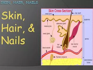
Anatomy • Epidermis • Stratum germinativum (basal cell layer) • Mitosis occurs here • Contains melanocytes, producing melanin • Stratum corneum • As cells rise, they die and their cytoplasm is converted to keratin, which has a rough, horny texture • This layer undergoes constant shedding • Dermis • Mostly connective tissue, primarily collagen • Provides support and nourishment of epidermis • Blood vessels, nerves, muscle, sweat glands, sebaceous glands, hair follicles • Subcutaneous Layer (Hypodermis) • Consists mostly of fat • Provides protection, insulation, and caloric source
Anatomy • Hair • Composed of keratin • Can be fine (vellus hair) or darker and thicker (terminal hair) • Sebaceous glands • Produce sebum through hair follicles, which make skin oily. Prevent water loss. • Sweat glands • Eccrine – smaller, coiled tubules which open to skin surface • Apocrine – larger, open to hair follicles. Located mainly in axillae and genital area. Produce thick secretions, which react with bacteria on skin surface to produce body odor • Nails • Composed of keratin • Clear with highly vascular bed of epithelial cells underneath Used to measures what? Pulse oxymetry!
Developmental Considerations • Infants • Lanugo – fine soft hair present at birth • Skin is thinner, less fat – more prone to dehydration and hypothermia • Pregnancy • Linea nigra – line down midline of abdomen • Chloasma – face of pregnancy • Striae gravidarum – stretch marks • Aging • Stratum corneum thins, loss of collagen, elastin, and fat, decrease of sebaceous and sweat glands, • More prone to dehydration and hypothermia Chloasma
History • History of skin disease • What was it? How was it treated? • Does it run in the family? • Significant familial predispositions – allergies, hay fever, psoriasis, eczema, acne • Any know allergies? • Any tattoos or birthmarks? • Use of nonsterile equipment for tattoos increases risk of Hep C • Change in pigmentation • Might suggest systemic illness (jaundice) • Change in a mole • Pruritus • Any dryness? Is it seasonal? • Xerosis – dry • Seborrhea - oily
History • Excessive bruising • Consider abuse • Frequent minor trauma may be sign of alcohol abuse • Rash or lesion • Onset • Location • Spread • Character or quality • Duration • Associative factors – pets, co-worker? • Alleviating and aggravating factors – what have you tried to do? • Patient’s perception - what do you think it is? • Medications • Prescription and over-the-counter • May indicate allergy to medication
History • Hair loss or growth • Gradual or sudden? • Hirsutism – unusual growth • Change in nails • Exposure to hazards • May be environmental or occupational • Bitten by bee, tick, mosquito? • Exposure to plants or animals? • Self care • What cosmetics, soaps, chemicals? • Possible allergies
General pigmentation – should be even throughout Benign pigmented areas Freckles (macules) on sun exposed skin Nevi (moles) Junctional nevi – macular only Compound nevi – macular and papular Dysplastic - precancerous Birthmarks Vitiligo – absence of melanin in patchy areas Physical Examination - Color ***** • ABCDE of malignant melanoma • Asymmetry – one lesion that is • not regularly round or oval • Border – irregular • Color – variations • Diameter – greater than 6mm • Elevation
Changes in Color in Light Skinned People • Pallor • Pale, white color caused by decrease of blood flow (vasoconstriction) or decrease in hemoglobin • Shock, anemia • Erythema • Redness due to increased blood flow (vasodilation) • Fever, inflammatory process, emotions, CO poisoning • Cyanosis • Bluish, purplish hue due to decreased perfusion of tissues • Hypoxemia due to heart failure, shock, chronic bronchitis • Jaundice • Yellow, orange hue due to jaundice (increased bilirubin in blood) • Due to liver problems such as hepatitis, cirrhosis
Color Changes in Darker Skinned People • Pallor • Brown skinned people will be more yellow. Black skinned people will be more gray • Palpebral conjunctiva and nail beds should be observed • Erythema • Cannot be observed • If fever suspected, check skin for warmth. If edema, check skin for tightness • Cyanosis • Darker skinned people have normal bluish tone on lips • Palms, but not clearly evident, other clinical signs should be observed • Jaundice • Hard and soft palate must be observed in addition to sclera of eyes • Dark urine also present Table 12.2
Skin Assessment (cont.) • Temperature • Check skin with dorsa of hands • Hyperthyroidism may cause increase of temp • Moisture • Diaphoresis may occur during fever or exercise • Dehydration can be observed by dry mucous membranes in mouth and cracked skin • Mobility and Turgor • Mobility is ease of skin rising when pinched. Turgor is returning back to its place • Slow turgor can be indicative of dehydration. “Tenting” if severe dehydration. • Lesions • A lesion is any traumatic or pathological change in skin • Describe using ABCDE, also noting location and exudate • Roll nodule gently between fingers to assess depth • Ultraviolet light is used if fungal infection suspected (Wood’s light)*****
Skin Assessment - shapes • Annular • Circular, beginning in center and spreading to periphery (ringworm) • Polycyclic • Annular lesions that grow together • Confluent • Lesions run together (hives) • Discrete • Individual lesions that remain separate
Shapes • Grouped • Clusters of lesions (contact dermatitis) • Gyrate • Twisted, coiled • Target • Concentric rings of color • Linear • Scratch like, stripe • Zosteriform • Follow nerve route (shingles)
Primary vs. Secondary • Primary skin lesions • Variations in color or texture that may be present at birth, such as moles or birthmarks, or that may be acquired during a person's lifetime, such as those associated with infectious diseases (e.g. warts, acne, or psoriasis), allergic reactions (e.g. hives or contact dermatitis), or environmental agents (e.g. sunburn, pressure, or temperature extremes). • Secondary skin lesions • Changes in the skin that result from primary skin lesions, either as a natural progression or as a result of a person manipulating (e.g. scratching or picking at) a primary lesion.
Primary Skin Lesions • Macule • color change and less than 1 cm • may be to darker or lighter • Freckles, flat nevi, hypopigmentation, petechiae • Patch • Color change and greater than 1cm • Mongolian spots, vitiligo, chloasma
Primary Skin Lesions • Papule • Elevated lesion less than 1cm in diameter • Due to elevation in epidermis • Ex: wart, elevated nevus • Plaque • Elevation greater than 1cm in diameter • Ex: psoriasis
Primary Skin Lesions • Nodule • Elevated solid greater than 1cm • Extending deeper into dermis • Tumor • Greater than few cm in diameter • May be firm or soft
Primary Skin Lesions • Wheal • Superficial, raised, transient, and erythematous lesion • Ex. Mosquito bite, allergic reaction
Primary Skin Lesions • Cyst • Encapsulated fluid filled cavity in dermis or subcutaneous layer • Vesicle • Elevated cavity containing free fluid, clear • Less than 1cm diameter • Ex: herpes simplex, varicella zoster
Primary Skin Lesions • Bulla • Larger than 1cm in diameter • Superficial in epidermis, thin walled • Ex: blisters, burns • Pustule • Pus in cavity • Ex: impetigo, acne
Secondary Skin Lesions • Crust • Thick, dry exudate after rupture or drying up of vesicle or pustule • Ex: Impetigo, scab following abrasion • Scale • Dry or greasy flakes of skin resulting from shedding of excess keratin cells • Ex: psoriasis, eczema, seborrheic dermatitis
Secondary Skin Lesions • Fissure • Linear cracks extending into dermis • Ulcer • Deep depression extending into dermis • May bleed. Leave scar. • Excoriation • Self inflicted abrasion often from scratching
Secondary Skin Lesions • Lichenification • Tightly packed papules from prolonged intense scratching • Keloid • Hypertrophic scar • Cannot be removed surgically • More common in black people
Skin Lesions associated with AIDS – Kaposi’s Sarcoma • Patch stage • Early lesions are faint and pink • Advanced stage • Widely disseminated lesions involving skin, mucous membranes, and visceral organs • Violet colored tumors on nose and face • Epidemic stage • Lesions develop into raised papules of thickened plaques. • Oval in shape and vary in color from red to brown.
Hair and Scalp • Ringworm may develop in scalp of school age children • Abnormalities in amounts and location of hair can be attributed to hormonal problems • Hirsutism – excess body hair • Observe for head or pubic lice, which are white ovals on hair shafts. • Dandruff is indicated by loose white flakes
Abnormal Conditions of Hair • Tinea capitis (scalp ringworm) • Lesions fluoresce blue-green under Wood’s light • Highly contagious • Toxic alopecia • Asymmetric balding that accompanies severe illness or chemotherapy • Regrowth after discontinuation of toxin
Abnormal Conditions of Hair • Folliculitis • Superficial infection of hair follicles • Multiple pustules • Furuncle and Abscess • Red, swollen, hard, tender, pus-filled lesion due to acute localized bacteria (staph) • Usually on back of neck, buttocks, wrists, or ankles • Furuncle is due to infected hair follicles • Abscess is due to traumatic introduction of bacteria into the skin. Deeper than furuncle
Nails • Good indicators of respiratory system health • Nail base • Normal is about 160° • Clubbing is the decrease of the angle of nail base (<160°) that occurs as a result of respiratory insufficiency, common in COPD (emphysema, chronic bronchitis) • In early clubbing, the angle actually increased to about 180° • Spongy nails Physiology of clubbing is not fully understood but respiratory insufficiency seems to dilate peripheral arteries, causing a round fingernail shape
Nails • Consistency • Variant thickness may suggest malnutrition • Thickening of nails is sign of arterial insufficiency • Color • Note any pigmentations – melanoma? • Cyanotic nail beds – poor peripheral circulation • Capillary refill • Indicator of peripheral circulation • Measured by depressing the nail bed until it is white and observing the time it takes for blood to return back to the nail • Normal time is less than 1-2 seconds and is indicated as “brisk.” “Sluggish” if greater than 2 seconds.
Developmental Considerations - Infants • Mongolian spots • Hyperpigmentation of sacrum, buttocks, abdomen, thighs, shoulders, or arms • Very common in blacks, Asians, and Native Americans • Should not be confused with abuse • Café au lait • “Coffee with milk” • Patches of hyperpigmentation • Normal
Developmental Considerations - Infants • Acrocyanosis • Bluish color around lips, hands, and feet • Usually is due to coolness and disappears after warming up • Persistent cyanosis is indicative of congenital heart disease • Cutis marmorata • Mottling of trunk and extremities due to coolness • If persistent, usually indicative of Down syndrome • Physiological jaundice • Common yellowing of skin in newborns, which usually appears after 4th day. UV light helps. • Carotenemia • Yellowing of skin due to ingestion of large amts of carotene.
Developmental Considerations - Adolescents • Acne • Most common skin problem • Acne occurs when the hair follicles, which are connected to sebaceous glands, become plugged with oil and dead skin cells. • Usually appear on face, shoulders, back, and chest • Can include papules, pustules, and nodules • Open comedones (blackheads) • Closed comedones (whiteheads)
Acne • Open comedones are a less severe form of acne
Vascular Lesions - Hemangiomas • Port-Wine Stain (Nevus Flammeus) • Flat macular patch of mature capillaries • Benign • Strawberry Mark (Immature hemangioma) • Raised bright red area • Usually disappears by age 7 • Cavernous Hemangioma
Developmental Considerations - Pregnancy • Striae • Linea nigra • Chloasma • Vascular spiders
Developmental Considerations - Aging • Senile lentigines • Liver spots – melanocyte clusters • Usually on hands and face • Seborrheic keratosis • Raised, thick, crusted “mole” • Dry skin is common • Acrochordons • Overgrowths of skin – normal • Frequently occur on back, eyelids, axillae
Developmental Considerations - Aging • Decreased turgor, tenting of skin occurs • Hair growth decreases, thins • Fungal infections of toenails
Pressure Ulcers • Stage I • A reddened area on the skin that, when pressed, is "non-blanchable" (does not turn white). This indicates that a pressure ulcer is starting to develop. • Stage II • The skin blisters or forms an open sore. The area around the sore may be red and irritated.
Pressure Ulcers • Stage III • The skin breakdown now looks like a crater where there is damage to the tissue below the skin. • Stage IV • The pressure ulcer has become so deep that there is damage to the muscle and bone, and sometimes tendons and joints.
Sensory Perception Activity Mobility Skin Moisture Friction and Shear Nutrition 1-4 with the exception of friction & shear subscale 1-3 Range 4-23 The lower the score the higher the risk Eighteen or less: high risk older adult Braden Scale
Question 1 • A nurse is reviewing the health care records of clients scheduled to be seen at the health care clinic. The nurse determines that which of the following individuals is at the greatest risk for development of an integumentary disorder? • An elderly female • An adolescent • An outdoor construction worker • A physical education teacher
Question 2 • A clinic nurse notes that the physician has documented a diagnosis of herpes zoster in a client’s chart. On the basis of an understanding of the cause of this disorder, the nurse would determine that this definitive diagnosis was made following which diagnostic test? • Skin biopsy • Wood’s light examination • Culture of the lesion • Patch test
Question 3 • A nurse is assessing for the presence of cyanosis in a dark-skinned client. The nurse understands that which body are would provide the best assessment? • Back of hands • Earlobes • Palms of hands • Sacrum
Question 4 • Which of the following clients would least likely be at risk for the development of skin breakdown? • A client who is unable to move about and is confined to bed • A client incontinent of urine and feces • A client with chronic nutritional deficiencies • A client with a lowered mental awareness
Question 5 • A nurse provides home care instructions to a client diagnosed with impetigo. Which of the following would not be a component of the teaching plan? • Continue with the antibiotics prescribed • Wash the client’s dishes separately from those of other household members • It is not necessary to separate the client’s linin and towels from those of other household members • Wash hands thoroughly and frequently throughout the day