Burn Injuries for Proper Care
Learn about the classification, treatment, and healing process of burn injuries. Understand skin changes, pain management, and complications associated with burns. Explore the importance of fluid management and healing time.

Burn Injuries for Proper Care
E N D
Presentation Transcript
Chapter 28 Care of Patients with Burns Marion Kreisel MSN, RN NU230 Adult Health 2 Fall 2011
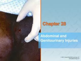
Pathophysiology of Burn Injury • Skin changes resulting from burn injury • Anatomic changes to largest Organ of the body • Functional changes: Protective Barrier against injury and microbial invasion, pain, excretory, • Temperature • ALL BURN INJURIES ARE PAINFUL! Third Degree Burn
CLASSIFICATION OF BURNS • According to the The American Burn Association (ABA) burns are classified as: • 1. Superficial-thickness wounds • 2. Partial-thickness wounds • a. superficial • b. deep • 3. Full-thickness wounds • 4. Deep full-thickness wounds • KNOW THIS
Superficial Thickness Burn 1. Least amount of damage 2. Only epidermis is injured 3. Usually caused by prolonged exposure to low intensity heat (sunburn) or short exposure to high heat (flash) 3. The degree of tissue damage is r/t agent, temperature, time of exposure, depth of skin eyes versus thigh 4. Redness, mild edema, pain, increased sensitivity to heat due to DESQUAMSTION ( peeling of dead skin) 5. Heals 3-5 days KNOW THIS SLIDE
Superficial Partial-Thickness Burn • 1. Involves the entire epidermis and varying depths of the dermis • 2. caused by heat injury to upper 1/3 of dermis leaving good blood flow. They are red moist and blanch. • 3. Small vessels bring blood to area and leak plasma, cause blisters. • 4. Pain increases, nerve endings exposed, and stimulation (touch or temperature changes) • 5. Heal in 10-21 days, usually no scar but pigment changes
Deep Partial-Thickness Burn • Extend deeper into skin dermis and kill more cells. Therefore no blisters b/c tissue is dead, thick and sticks to the underlying dermis. Does not remove easily. • Red, dry, white areas b/c damage to blood vessels. • Moderate Edema, less pain b/c nerve endings are destroyed. • TX: HYDRATION, nutrition and O2 for regrowth of skin cells and prevent conversion to deeper burns • Heal in 3-6 weeks with scar formation
Full-Thickness Burn • Destruction of the entire epidermis and dermis leaving no cells to regenerate. • 2. What ever doesn’t heal and close need wound graft. • They are hard, dry, leathery (eschar: dead tissue that must slough off or be removed before healing can occur. Very difficult to remove. • Lots of edema under the eschar and if wound is circumferential tightness happens • May have to have Escharotomies (incsion through eschar) or Fasciotomies (incision through eschar and fascia). • Wounds avascular, reduced sensation • Healing Time weeks to months
Deep Full-Thickness Burn • Extend beyond the skin into underlying fascia and tissue. • 2. damage muscle, bone, tendons and leave them exposed. • Occur from flame, electrical or chemical exposure • The wound is black and depressed and loss of all sensation. • TX: early excision and grafting. Amputation may be needed.
Vascular Changes Resulting from Burn Injuries • Fluid shift—third spacing or capillary leak syndrome, usually occurs in the first 12 hr and can continue 24 to 36 hr. It is a continous leak of plasma from the vascular space -> loss of blood volume ->decrease B/P • Profound imbalance of fluid, electrolyte, and acid-base, hyperkalemia and hyponatremia levels, and hemoconcentration (due to dehydration), severe edema • Fluid remobilization after 24 hr,when capilary leakage stops. diuretic stage begins 48 to 72 hr after injury b/c fluid shifts back into vascular system, hyponatremia and hypokalemia
Changes Resulting from Burn Injury • Changes include: • Cardiac: Increase HR, Decrease CO and then shift with fluid resusitation • Pulmonary: • GI (Curling’s ulcer): Acute gastroduodenal ulcer r/t stress of burns. Decrease GI blood flow and increase mucosal damage. • Metabolic: hypermetabolism which causes increase in O2 need and calories • Immunologic: destroys protective barrier of the skin, increase of infection
Compensatory Responses to Burn Injury • There are 2 responses: • Inflammatory compensation: can trigger healing however causes blood vessels to leak fluid into the interstitial space and WBC to release chemicals that trigger local reactions. Helpful in the beginning short term. • Sympathetic nervous system compensation: occurs when any physical or psychological stressors are present.
Etiology of Burn Injury • Dry heat: open flame, most common, brief exposure high temperature • Moist heat: hot liquids and steam. Scalding injuries • Contact burns: hot metal, tar, grease leads to full-thickness injury. Space heater, iron, molten metals, • Chemical injury: skin contact, inhaled, swallowed. Severity of injury depends on duration of contact, concentration, , amount, and action of chemical • Electrical injury: Burns when electrical current enters body. Small looking outside large damage inside • Radiation injury: Exposure to large doses of radioactive material
Electrical Burn The mechanism of electrical injury: Currents passing through the body follow the path of least resistance to the ground
Emergent Phase of Burn Injury • First phase, or emergent phase, continues for about 48 hr. • Goals of management include: • Secure airway • Support circulation—fluid replacement • Prevent infection • Maintain body temperature • Provide emotional support
Injuries to the Respiratory System • Direct airway injury • Wheezing, stridor, hoarness. If these noises stop it is an RESPIRATORY EMERGENCY! • Carbon monoxide poisoning • Thermal injury • Smoke poisoning • Pulmonary fluid overload • External factors
Cardiovascular Assessment • Hypovolemic shock is a common cause of death in the emergent phase in patients with serious injuries. • Monitor vital signs. • Monitor cardiac status, especially in cases of electrical burn injuries.
Renal/Urinary Assessment • Changes are related to cellular debris and decreased renal blood flow. • Myoglobin is released from damaged muscle and circulates to the kidney. • Assess renal function, blood urea nitrogen, serum creatinine, and serum sodium levels. • Examine urine for color, odor, and presence of particles or foam.
Skin Assessment • Determine size and depth of injury. • Determine percentage of total body surface area affected. • Use "rule of nines," using multiples of 9% of total body surface area.
Gastrointestinal Assessment • Changes in GI function are expected. • Decreased blood flow and sympathetic stimulation during the emergent phase cause reduced GI motility and paralytic ileus. • Assess for GI bleeding.
Burns: Nonsurgical Management • IV fluids • Monitoring patient response to fluid therapy • Drug therapy:
Burns: Surgical Management • Escharotomy • Fasciotomy
Acute Phase of Burn Injury • Begins about 36 to 48 hr after injury and lasts until wound closure is completed • Care directed toward continued assessment and maintenance of the cardiovascular and respiratory systems, as well as toward GI and nutritional status, burn wound care, pain control, and psychosocial interventions
Assessment • Assessments include those of: • Cardiopulmonary • Neuroendocrine • Immune • Musculoskeletal
Nonsurgical Management: Acute Phase • Mechanical débridement: • Hydrotherapy • Enzymatic débridement: • Autolysis • Collagenase
Dressing the Burn Wound 1. Standard wound dressings: gauze, ointment 2. Biologic dressings: Used temporary for closure; Skin membranes when applied rapidly adheres and promotes healing or prepares skin for graft. • Homograft—human skin • Heterograft—skin from other species 3. Amniotic membrane: Large, low cost, high availability, very successful. Full thickness adheres to wound in partial thickness works as a dressing, frequent changes b/c no blood supply and disintegrates. 4. Cultured skin: Take cells from body and grow in lab
Dressing the Burn Wound(Cont’d) • 5. Artificial skin: Silastic epidermis and porous dermis from beef collagen and shark cartilage • 6. Biosynthetic dressings: made from biosynthetic and synthetic materials • 7. Synthetic dressings: made of solid silicone and plastic membranes. Transparent so do not have to keep removing to check site, promote healing • 8. Pressure dressings are applied after the graft heals to help prevent contractures and tight hypertrophic scars, which can inhibit mobility. For best effectiveness, pressure garments must be worn at least 23 hours a day, every day, until the scar tissue is mature (12 to 24 months).
Surgical Management • Surgical excision • Wound covering: • Skin graft
Meshed Autograft Application Healing
Nonsurgical Management • Drug therapy: silver sulfadiazine (Silvadene) Watch for allergic reaction and decrease in WBC • Isolation therapy • Environmental management
Rehabilitative Phase of Burn Injury • Rehabilitation begins with wound closure and ends when the patient returns to the highest possible level of functioning. • Emphasis during this phase is on psychosocial adjustment, prevention of scars and contractures, and resumption of preburn activity.
Rehabilitative Phase of Burn Injury (Cont’d) • This phase may last years or even a lifetime if patient needs to adjust to permanent limitations.
Chapter 28 NCLEX TIME Care of Patients with Burns
Question 1 What is the estimated number of fire- and burn-related deaths that occur yearly? • 2000 • 4000 • 6000 • 10,000
Question 2 After smelling smoke, nurses find a patient who is in bed with his leg in traction beating small flames on his clothing with a pillow. He is coughing and gasping for air. One nurse activates the hospital emergency call system for fire, and the other nurse will perform which action first? • Administer oxygen to the patient. • Assess for airway patency. • Smother the flames. • Obtain vital signs.
Question 3 A patient who has suffered extensive burns over his left leg has had skin grafts and is now being prepared to wear a pressure dressing over his leg. The patient asks how long he will need to wear this dressing. The best answer would be: • “Until the swelling in your leg is gone.” • “For the next 12 months, but only while you are awake, until the scar tissue has healed.” • “During the night hours, when you are in bed, until the scar tissue heals.” • “You will need to wear this for 23 hours a day for the next 12 to 24 months, until the scar tissue has matured.”
Question 4 It has been 12 hours since a patient has been admitted for burn and inhalation injuries. He had been wheezing audibly, but at this time the nurse notes that his wheezing has stopped. The nurse should: • Document this improvement in the patient’s condition. • Re-assess his breathing in an hour. • Check the patient’s Spo2 level. • Notifiy the physician immediately
Question 5 A patient has been receiving dressing changes with silver sulfadiazine (Silvadene) for burn injuries over both lower arms. The nurse notices that the patient’s white blood cell count has dropped significantly over the past 4 days. This change may indicate: • The patient’s infection is improving • An allergic reaction to the silver sulfadiazine • Kidney disease • An electrolyte imbalance