Download
1 / 44
480 likes | 773 Views
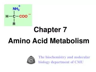
Chapter 10 Amino acid & Protein Analysis Qijun Wang 2005-4-12. Chapter 10-1 Amino Acids Analysis. What is amino acid?. What is amino acid? Amino Acid: aminated carboxylic acid (R-COOH). R group. Examples. -amino acetic acid. -amino propanoic acid. , -2 amino caproic acid.

E N D
What is amino acid? What is amino acid? Amino Acid: aminated carboxylic acid (R-COOH) R group
Examples -amino acetic acid -amino propanoic acid ,-2 amino caproic acid
Classification of Amino Acid 1. By the location of Amino-group : / / -AA 2. By its acidity: neutral/acidic/ basic AA ratio of Amino-group to carboxylic group 3. By whether containing phenyl group aromatic / non aromatic AA 4. By its occurrence in protein Protein / non protein AA 5. By polarity of R group : polar / apolar side chain AA 6. By its nutrient value to human: EssentialAA and non-essential AA
Physical characteristics • ALL the pure -AAs are Colorless crystal , with quite high melting point (>200 deg.C) and water solubility,
HCL NaOH H AA are ampholyte Anion, when at high pH Cation, when at low pH zwitterion, when at isoelectric point (pI) NOTE: peptide or protein also have both acid and base properties. They share the same property of being positively charged at low pH and negatively charged at high pH.
+ - - + - + - + - + - + - + - + - + - Isolectric Point (pI) of AAs anode cathode Zwitterion in pI sol’n. No move to either of Electrode. And with Lowest solubility Cation in acidic sol’n. Anion in basic sol’n. pH溶液<pI pH溶液>pI
Note on pI • The Acidity of neutral AA is stronger than its Basicity, which means the dissociation degree of reaction (i) is larger that of reaction(ii). Therefore the pH of neutral AA water solution is less than 7 。 H2N-CHR-COOH +H2O H2N-CHR-COO- +H3O+ (i) H2N-CHR-COOH +H2O H3N+-CHR-COOH +OH- (ii) * therefore, [anion] >[cation], to get pI, more acid is to be added. • pI of neutral AAs is around 5.6~6.3, • pI of acidic AAs is around 2.8~3.2; • pI of basic AAs is around 7.6~10.8。
Ninhydrin Reaction Note: 1, Ninhydrin solution is made in basic solution of phosphate buffer (pH8.04). 2, The reaction products of all AAs except proline, are purpule-bule(540nm), and for proline is yellow(440nm) 3, protein and peptide also can occur this reaction due to their containing amino group. 4. This reaction can be used to quantitative and qualitative analysis, with the help of spectrophotometer, TLC.
Automatic AA analyzer From pH2.2 to pH 6.4
Separation principles H+-type ion exchange Resin: consists of relatively chemically inert polymer, which has quite strong acidic side-chain constituents such as –SO3H,-CH2SO3H *suitable for A/B/Neutral conditions - + Separate aspartic acid and lysine by cation exchange resin.
Automatic AA analyzer Absorbent: cation ion exchange resin, Eluting sol’n: citric acid buffer of pH2.2,pH3.3,pH4.0 and pH6.4 Extracted and evaporated AAs is required to be dissolved in pH2.2 citric acid. Eluting order: acidic AAs,polar AAs, apolar AAs, and basic. For AAs in a same catalog, low mass AA is eluted out first.
Aromatic AAs Aromatic AAs absorb light in the near ultraviolet (230-300nm). phenylalanine tyrosine Note: This UV absorption property of protein is solely determined by the content of these 3 aromatic AAs. However, far ultraviolet (190nm)absorption of protein stems from the peptide bond. tryptophan
AAs assay by GC Principle: Non-volatile AA Volatile AA-derivate
Protein Analysis What is protein: • polymer of 20 - amino acids, with mol.wt from 5000 to1000,000 daltons. • N is most distinguished element: among the composing elements of C,H, N, O, S, for some proteins: P, Cu, Fe, I. • N content in different proteins ringing from 13.4% -19.1%, and averagely 16%. • Therefore protein coefficient is 6.25 for most proteins. 5.70 is only for wheat and its products proteins according to AOAC method. • Most abundant component in cells: 50% of dry cells by weight
Protein content in food (%) Cereals: (%) Brown Rice 7.9 Polished rice 7.1 Wheat flour, whole-grain 13.7 Corn flour, whole-grain 6.9 Corn starch 0.3 legumes: Soybean, raw 36.5 Beans, kidney, raw 23.6 Tofu, raw, regular 8.1 Fruits & vegetables: Apple, raw, with skin 0.2 Strawberry, raw 0.6 lettuce , raw 1.0 Meat, poultry, fish: Beef 18.5 Dry beef 29.1 Chicken, breast meat, raw 23.1 Ham 17.6 Egg, raw, whole 12.5 Finfish, raw, 17.9 Dairy products: Milk, whole, fluid 3.3 Milk, skim, dry 36.2 Cheese, cheddar 24.9 Yogurt 5.3 * High quality protein ?
Conversion factors for Foods N to Protein conversion factors
Proteins functions NO proteins no life! 1, Structural proteins: Such as keratin; myosin, actin; glycoprotein, lipoprotein, 2, biological active proteins: Such as: Enzymes, hemoglobin, myoglobin, ferritin, antibody, glycoprotein, lipoprotein
Classification 1. According to whether containing non-proteins components : • Simple protein: only containing AAs upon hydrolysis, such as Egg Albumin; myosin, actin, insulin; • Conjugated protein: AAs + non-AAs upon hydrolysis; Such as lipoprotein; glycoprotein; hemoglobin, ferritin; majority of enzymes 2. According to theirs solubility: • Non-water soluble protein: filament protein: myosin, actin, keratin • Water soluble proteins: hydrophilic groups outsides (apolar groups), and hydrophobic groups (-OH, -SH, -COOH,-NH2) insides, most global proteins, enzymes. * Enzymes working conditions: mild conditions。
Main Properties of Protein 1. As polymer of AAs, proteins have both acid and base properties.zwitterion & pI& electrophoresis. 2. Most proteins are water soluble, and unable pass throughdialysis membrane. 3. Denaturation: denaturants such as heat, acid, alkali, salt, detergents can altered solubility and functional properties of proteins. reversible/non-reversible denaturation. 4. Ultraviolet absorption at 280 nm, due to the presence of 3 aromatic acids residues, i.e. tyrosine, tryptophan, phenylalanine.
Kjeldahl’s method Principles: 1. Digest the organic compounds with strong sulfuric acid in the presence of catalysts while heating. 2. The total organic N is converted to ammonium sulphate. 3. Neutralize the digested sol’n with abundant alkali. Here, the N is converted to ammonium hydroxide, and then being distilled into a boric acid solution and converted to ammonium borate. 4. Titrate ammonium borate with strong acid. (please notice that N: HCl = 1:1) 5. N content in proteins is averagely 16%.
Principles of Kjeldahl’s method 1. Digestion NCOC + H2SO4 (NH4)2SO4 + CO2 + SO2 + H2O 2. Neutralization &distillation 2NaOH +(NH4)2SO4 2NH3↑+Na2SO4 + 2H2O 3. Absorption by boric acid : 2NH3 + 4H3BO3(NH4)2B4O7 + 5H2O 4. Titration by strong acid (NH4)2B4O7 + 5H2O + 2HCl 2NH4Cl + 4H3BO3 (*NCOC,N containing organic compounds, N:HCl = 1:1) 催化剂 煮沸
Apparatus used in Kjeldahl I. Digestion apparatus II. Distillation & absorption apparatus (II) (I)
Points that need your close attention 1. Amount of protein sample and reagents used should be proportional. 2. All the working solution should be prepared with ammonia-free distilled water 3. Mildly heating When digestion, so that no sample to spatter onto flask wall. 4. Rotate the flask while digestion. 5. Add antifoam(silica oil) if necessary. 6. 30% hydrogen peroxide can accelerate the digestion. 7. At the end of fully digestion, the solution should be clear light-blue or greenish.
Points that need your close attention 8. Digestion should be carried out in a ventilating cabinet. 9. Connect well the distillation apparatus before adding alkali into digested solution. 10. Add abundant alkali until there are red copper hydroxide formed. 11. Absorption solution should be less than 40 deg.C throughout the absorption. Cold water bath is a good choice to lower the temp. 12. Using indicating paper to help for the determination of distillation terminus. 13. Indicators of methylene blue and methyl red are added to absorption bottle before carrying on the distillation.
Micro Kjeldahl Method Notes: 1. Applicable to all types of foods; 2. accurate, as an official method for crude protein content.3. poorer precision than the biuret method.
The Biuret Method Principle: This reaction is characterized by the development of a purple coloration from the complexing of cupric ions with peptide bonds in an alkaline medium. The wavelength , however, varies with the nature of the protein: from 540 to 650 nm, often at 550 nm. Q:1. Is this suitable for AAs? 2. what is Biuret Reaction?
Application and advantages • Applications: Suitable for cereals, meat, soybean, and isolated proteins. But not for milk, as reducing sugars (lactose) can reduce copper ion. • Advangtages: 1. less expensive than Kjeldahl method, rapid, simplest methods for protein analysis. 2. color deviations are encountered less frequently than with Folin-Lowry, UV absorption, or turbidimetric methods. 3. very few substances other than proteins in foods interfere with the biuret reaction. 4. Detect N only from the peptide and protein sources. Disadvantages: • Not very sensitive as compared to the Folin-Lowry Method. Require at least 2-4 mg protein for assay. • Bile pigments, high conc of ammonium salts interferes with the reaction. • Color varies with different proteins. Gelatin gives a pinkish-purple color. • Not an absolute method: color must be standardized against known proteins(e.g., BSA) • * Introduction of 30% isopropyl alcohol can reduce the reaction time from 35 to 10 mins. You also can try “Heating”.
Dumas (N combustion) • Principle: Samples are combusted at high temp (700-1000 deg.C). The N released is quantitated by GC using a thermal conductivity detector (TCD). • Procedure: Samples (100-500 mg) are weighed into a tin capsule and introduced to a combustion reactor in automated equipment. The N released is measured by a built-in GC. • Advantages: 1, Applicable for All proteins; 2, No hazardous chemicals; 3, Saving time: within 3 mins; 4, High performance: recent automated instruments can analyze up to 150 samples without attention. • Disadvantages: Measures total organic nitrogen, not just Protein N.
The lowry’s Method (Folin-phenol method) Principle: • Folin reagent (phosphomolybdic and phosphotungstic acid)is reduced to a blue molybdenum complex, mainly by the phenolic groups of tryptophan and tyrosine. 2. Lowry greatly increased the sensitivity of the determination by preceding the reaction by pretreatment with a copper reagent in a basic medium. (Mistake in Sol’n B at p163)
Procedures of lowry’s Method 1. Dilute protein sample to contain 20-100 ug. 2. K Na tartrate-Na2CO3 solution is added after cooling and incubated at RT for 10 min. 3. CuSO4- K Na tartrate-NaOH solution is added after cooling and incubated at RT for 10 min. 4. Freshly prepared Folin Reagent is added, then the reaction mixture is mixed and incubated at 50 deg.C for 10 min. 5. Absorbance is read at 650 nm. 6. A standard curve of BSA is carefully constructed for estimating protein conc of the unknown.
Properties of lowry’s Method • Applications: Widely used in protein biochemstry, because of its simplicity and sensitivity. But not widely used in Food proteins analysis without extracting proteins from the food mixtrue. Advantages: 1. 50-100 times more sensitive than biuret method, and 10-20 times than 280 nm UV absorption method. 2. Less affected by turbidity of the sample. 3. More specific than most other method. 4. Relatively simple, can be done in 1-1.5 hours. Disadvantages: 1. Color varies with different proteins to a greater extent than the biruet method. 2. Color is not strictly proportional to protein conc. 3. The reaction is Interfered with to a varying degree by sucrose, lipids, phosphate buffers, monosaccharides, and hexoamines. 4. high conc., of reducing sugars, ammonium salfate, and sulfhydryl compounds interfere with the reaction.
UV 280 nm absorption Method Principle: 1. Proteins show strong absorption at UV 280 nm, primarily due to aromatic amino acids of tryptophan and tyrosine residues in proteins. 2. The content of Try and Tyr in proteins from each food sources is fairly constant. Thereby, the extinction coefficient(E280) or molar absorptivity (Em) must be determined for individual proteins for protein content estimation. Applications: 1. Used for protein content of meat and milk product. This technique is better applied in a purified protein system or to proteins that have been extracted in alkali or denaturing agents such as 8 M urea. Advantages: 1. Rapid; 2. Non interference from ammonium sulfate. 3. Non destructive. Disadvantages: 1. aromatic amino acids contents in proteins from various food sources differs considerably!
Coomassie Blue staining procedures 1. Reagents Coomassie Blue R-250 Methanol Glacial acetic acid 2. Gel Stainning solution, 1 L 1.0 g Commassie Blue R-250 450 ml water 450 ml methanol 100 ml Glacial acetic acid 3. Gel destaining solution, 1 L 100 ml Methanol 100 ml Glacial acetic acid 800 ml Water 4. Staining Procedure 1. Pick up the gel into (20 ml, usually enough) Staining solution in a container and agitate for 10 min for 0.75 mm Gel and 20 min and 1.5 mm gel. The staining solution can be reused several times. 2. Take the gel out and rinse the gel with a few changes of water in a new container 3. Add 50 ml destaining solution. Strong bands are visiable immediately on a light box, and 1 hour usually is enough. 4. To destain completely, change destaining solution 2-3 times and agitate overnight. 5. Scan or take photo to record the result.
Two dimensional Electrophoresis 2-DE: the technique to separate proteins in the first dimension according to their isoelectric point, by Isoelectric Focusing(IEF), and in the second dimension according to their molecular weight, by SDS-PAGE. 2-DE combined with protein identification basing on microsequencing, amino acid composition and Mass spectrometry, provides an invaluable tool for proteomic studies. Step 1 — Sample Prep Step 2 — First-Dimension (IEF) Separation Step 3 — Second-Dimension (SDS-PAGE) Separation Step 4 — Protein Detection by Staining /Destaining