Download
1 / 28
300 likes | 550 Views
Initial management of PUV. Ahmed Al-Sayyad MD,FRCSC. Clinical Presentation. Boys with PUV can present with a variety of symptoms and at various ages They range from newborns with life-threatening renal and pulmonary conditions to older children with minor voiding dysfunction

E N D
Initial management of PUV Ahmed Al-Sayyad MD,FRCSC
Clinical Presentation • Boys with PUV can present with a variety of symptoms and at various ages • They range from newborns with life-threatening renal and pulmonary conditions to older children with minor voiding dysfunction • In general, the symptoms are age dependent; the more severely affected boys present earlier in life • Antenatal hydronephrosis diagnosed with prenatal ultrasonography is the most common presentation
Clinical Presentation • Obstruction leads to decreased fetal urine output and results in oligohydramnios • The observation of marked hydro, a distended bladder, and a thickened bladder wall in utero strongly supports the diagnosis of valves • Neonates with sever PUV can present with Pulmonary Hypoplasia , intrauterine growth retardation, failure to thrive, lethargy, and poor feeding • Older children can present with urinary tract infection, voiding dysfunction or renal insufficiency
Approach • Hx :Pre and postnatal • Exam: Abdomen,genitalia and back • Blood work: CBC,lytes,Cr,?acid base • Bladder drainage • Abx prophylaxis • US • VCUG • Careful monitoring
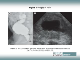
US • Hydroureteronephrosis • Increased echogenicity • Peri-renal urinoma • Thickened bladder
VCUG • Bladder is thickened and trabeculated, diverticula may be present, bladder neck is elevated and the proximal urethra is dilated, and the actual valve structure is often visible • Vesicoureteral reflux is present in at least 50% of valve patients at the time of diagnosis • The incidence of reflux has been found to be higher in neonates than in older children • There is an 80% incidence of reflux on the left side in patients with unilateral reflux for no apparent reason
Investigations • Initial laboratory evaluation of the newborn with valves is usually misleading because of the effects of maternal renal function mediated through the placenta • It will take at least 48 hours for the serum levels of creatinine and blood urea nitrogen to accurately represent the child's intrinsic renal function • Creatinine, blood urea nitrogen, and electrolyte values should be determined twice daily for the first few days of life until they plateau
Bladder Drainage • Initial management of all patients with PUV requires immediate bladder drainage • This should be performed even if the diagnosis has not been confirmed by VCUG • Neonates can be catheterized with a 3.5 or 5 French pediatric feeding tube • Foley catheters have been used with success, but there have also been reports that the balloon causes irritation and resultant bladder spasms • After successful initial bladder drainage and when the patient's medical condition has stabilized, the next step is to permanently destroy the valves
Valve Ablation • Transurethral valve ablation is the 1st treatment choice • A Bugbee electrode or a pediatric resectoscope with a hook or cold knife can be used to incise the valves • A number of authors report use of a cystoscope and laser to disrupt valves • Some surgeons prefer incision at 12-o'clock position; others prefer incisions at 4- and 8-o'clock, and others all three • Although most valves are thin and do not bleed at surgery, it is preferable to leave a catheter in place for 24 hours after incision • The valve remnants resolve after incision, and there is often no evidence of them on later cystoscopic examination
Cutaneous Vesicostomy • If the infant is too small for safe instrumentation for valve ablation, a cutaneous vesicostomy can be performed as a temporary measure • The vesicostomy provides adequate drainage of the upper tracts in more than 90% of cases • There has been concern that vesicostomy would cause permanent loss of bladder volume, but this has not proved to be true, and vesicostomy does not significantly affect bladder capacity
Upper Tract Diversion • There is controversy about the superiority of upper tract diversion vs. vesicostomy regarding long-term results and measured renal function, bladder function, and somatic growth in each group • the current consensus is that neither initial treatment is superior in promoting renal function and somatic growth • The current consensus is that both approaches eventually yield similar results and that infants who undergo initial upper tract diversion are at the disadvantage of needing more surgical procedures • Today, upper tract diversion is usually limited to those patients who fail to respond to bladder-level drainage • Upper tract diversion Is considered if bladder-level drainage is insufficient to prevent infection or to drain the upper tracts adequately
Upper Tract Diversion • If the serum creatinine concentration drops below 2.0 mg/dL (150 μmol/L), it is safe to rely on improved bladder drainage for additional kidney improvement • If the creatinine concentration remains above 2.0 mg/dL (150 μmol/L) after 10 days of adequate bladder decompression and if hydronephrosis is unimproved, upper tract diversion may be considered • The type of diversion remains the surgeon's choice, options are high loop ureterostomy, ring ureterostomy, pyelostomy, and end ureterostomy • If upper tract diversion is performed, reconstructive surgery to internalize the urinary tract should be delayed until the bladder and upper tracts have improved as much as can be expected
Management of VUR • Reflux in PUV is considered secondary to bladder outlet obstruction • the initial management of reflux is relief of obstruction • Reflux resolves after valve ablation in between 20% and 32% of refluxing ureters • Most reflux resolves within several months, but some can take as long as 3 years • Reflux is more likely to resolve when it is associated with a better functioning kidney
Management of VUR • Children with initial bilateral reflux are more likely to have reflux resolve than are those with unilateral reflux • As for any child with vesicoureteral reflux, they must be maintained on prophylactic antibiotics to prevent infection • If persistent high-grade reflux is a clinical problem because of urinary tract infections or incontinence, bladder function and drainage must be reviewed • Inadequate emptying and high storage pressures are the usual causes of persistent reflux
Management of Hydronephrosis • Nonrefluxing hydronephrosis resolves in 49% of patients and may do so rapidly after valve ablation • This leaves a significant population of valve patients with persistent hydronephrosis for years despite adequate bladder emptying • The majority of patients with persistent hydronephrosis do not have obstruction at either the bladder outlet or the ureterovesical junction
Summary • PUV is one of the trickiest conditions to treat • It requires a very careful initial and long-term management • Our goal is to preserve all remnant kidney function by relieving obstruction and preventing infection • Special attention and long-term bladder management is a key in treating those patients