Elbow evaluation
Elbow evaluation. Part Two. Overview. Patient history Observation Palpation Bony Soft tissue Active/passive ROM Special tests Neurologic Sensory Motor DTRs Circulatory. Patient History. MOI What was the mechanism of injury? Describe or demonstrate.

Elbow evaluation
E N D
Presentation Transcript
Elbow evaluation Part Two
Overview • Patient history • Observation • Palpation • Bony • Soft tissue • Active/passive ROM • Special tests • Neurologic • Sensory • Motor • DTRs • Circulatory
Patient History • MOI • What was the mechanism of injury? Describe or demonstrate. • What were you doing when the injury occurred? Was it the result of throwing or swinging? • Did you receive a direct blow? • What was angle of impact? • What was the position of arm at impact? • Did it involve the neck or shoulder? Is there any pain in the shoulder or neck? • Was the wrist forced beyond its normal ROM? • Did you have a violent muscular contraction? • Did you hear or feel anything at the time of injury?
Patient History • Signs & Symptoms • Describe the symptoms? • Describe the pain. • Was it gradual or sudden onset? • Is it sharp or dull? Localized or diffuse? • Is the pain radiating down your arm? • Is the pain severe? Does it keep you awake at night? • Demonstrate what causes pain. • Do you feel any numbness, tingling, or burning? • Do you feel any weakness? • Does the arm feel tight or locked?
Patient History • Previous injury • Have you had a previous injury? • Did you see a physician? • What was diagnosis? • Were you fully recovered? • Did you rehab? If so, what exercises?
Observation • Overall position of person • Are they using the involved limb? • Is the arm in an abnormal position? • Is the limb hanging limp? • Do the shoulders look symmetrical? • Do there appear to be any deformities? • Swelling • Atrophy • Abnormalities
Observation • Skin color • Skin temperature • Any signs of trauma? • Carrying angle • Spasm
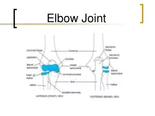
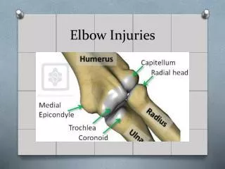
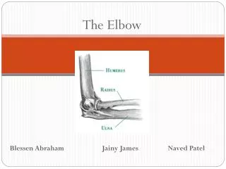
Palpation: Bone • Humerus • Medial epicondyle • Lateral epicondyle • Trochlea • Capitulum • Radius • Radial process • Radial tuberosity • Ulna • Olecranon process • Olecranonfossa • Ulnarstyloid • Cubitalfossa (tunnel) • Bony triangle
Palpation: Soft tissue • Olecranon bursa • Ulnar nerve • Radial nerve • Median nerve • Lateral collateral ligament
Palpation: Soft tissue • Medial collateral ligament • Annular ligament • Supracondylar lymph nodes • Biceps tendon • Triceps tendon
Palpation: Soft tissue • Biceps brachii • Triceps brachii • Brachialis • Brachioradialis • Anconeus • Pronatorteres • Pronatorquadratus • Supinator • Flexor carpiradialis • Palmaris longus • Flexor carpiulnaris • Extensor carpiradialis
Active/Passive ROM • Elbow: • Flexion (140-150) • Extension (0-10) • Wrist: • Flexion (80-90) • Extension (70-90) • Radial deviation (15) • Ulnardeviation (30-45) • Pronation (80-90) • Supination (90)
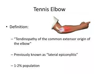
Special Tests • Forearm Compression Test (fracture) • Transverse Stress Test (fracture) • Percussion Test (fracture) • Longitudinal Stress Test (fracture) • Collateral Stress Test (MCL/LCL laxity) • Hyperextension Test (Anterior capsule) • Elbow Flexion Test (Cubital tunnel or ulnar nerve) • Tinel's Sign (at elbow) (ulnar nerve) • Milking Sign (MCL instability) • Cozen’s Resistive Tennis Elbow Test (tendinitis) • Resistive Tennis Elbow Test (tendinitis) • Passive Tennis Elbow Test (tendinitis) • Golfer’s Elbow Test (tendinitis)
Forearm Compression Test (fracture) • The examiner uses both of his or her hands to squeeze the pt’s radius and ulna together, being certain to test proximally, intermediately, and distally. • Pain indicates a positive test for forearm long bone fracture (radius or ulna).
Transverse Stress Test (fracture) • The examiner grasps the pt’s radius and attempts to translate it distally and proximally, effectively stressing the bone along its long axis. • The test should be repeated with the ulna. • Obvious sharp pain indicates a positive test for forearm long bone fracture.
Percussion Test (fracture) • The examiner simply percusses the bones of the forearm, paying special attention to bony landmarks. • This should include the radius, ulna, olecranon process, and the medial and lateral epicondyles. • Sharp pain indicates a positive test for forearm long bone fracture.
Longitudinal Stress Test (fracture) • The examiner first stabilizes the pt’s elbow, then grasps the radius and ulna individually, using both hands. • The examiner next pulls upwardly with one hand while pushing downwardly with the other hand, repeating several times. • Pain indicates a positive test for forearm long bone fracture.
Collateral Stress Test (MCL/LCL laxity) • The examiner begins by stabilizing the pt’s elbow with one hand and grasping the pt’s distal forearm with the other hand. • The examiner then applies a varus force with the pt’s elbow in 90 degrees of flexion, then 30 degrees of flexion, and finally in full extension. • A varus force tests the lateral collateral ligament of the elbow. • The examiner then repeats the procedure with the application of a valgus force. • A valgus forces serves to test the medial collateral ligament, which is normally taut in full extension, lax between 10 and 60 degrees of flexion, and taut at 90 degrees of flexion. • Laxity or instability indicates a positive test.
Hyperextension Test (Anterior capsule) • Examiner supports pt in passive endrange extension. • Elbow extension beyond 0 degrees with pain may indicate an injury to the anterior capsule.
Elbow Flexion Test (Cubital tunnel or ulnar nerve) • The pt simply actively holds the elbow in full flexion for 3-5 minutes. • A positive test results if the pt notes tingling or paresthesia in the ulnar distribution in the forearm and hand. • A positive test may indicate possible cubital tunnel syndrome and/or ulnar neuritis.
Tinel's Sign (at elbow) (ulnar nerve) • The examiner first palpates the pt’s cubital tunnel near the medial epicondyle. • The examiner then taps over the cubital tunnel, looking for some type of paresthesia from the ulnar nerve’s distribution. • Shooting pain down the forearm into the ring and fifth fingers indicates a positive test for ulnar neuritis.
Milking Sign (MCL instability) • The examiner prepositions the pt in 90 degrees of elbow flexion and shoulder abduction • The examiner then grasps the pt’s thumb and passively forces the pt’s shoulder into endrange external rotation • Pain at the medial elbow is considered a positive test for medial collateral ligament injury
Cozen’s Resistive Tennis Elbow Test (tendinitis) • Pt is sitting while examiner stabilizes the involved elbow while palpates along the lateral epicondyle. • With closed fist, the pt pronates and radially deviates the forearm and extends the wrist against the examiner's resistance • Pain along the lateral epicondyle region of the humerus or objective muscle weakness as a result of complaints of discomfort indicate a positive test for lateral epicondylitis
Resistive Tennis Elbow Test (tendinitis) • With the pt sitting, the examiner stabilizes the involved elbow with one hand and places the other on the dorsal aspect of the pt’s 3rd digit • Pt extends the third digit against resistance • Pain or weakness may indicate lateral epicondylitis
Passive Tennis Elbow Test (tendinitis) • With the pt seated, examiner passively pronates the forearm and flexes the pt’s wrist. • Pain on or near the lateral epicondyle may indicate lateral epicondylitis.
Golfer’s Elbow Test (tendinitis) • Pt is sitting or standing and makes a fist with the involved side • Examiner faces pt and palpates along the medial epicondyle with one hand and grasps pt's wrist with the other hand • Examiner passively supinates the forearm and extends the elbow, wrist and fingers • Pain along the medial aspect of the elbow indicates a positive sign for medial epicondylitis
Neurologic Exam: Sensory • Sensory dermatomes • C2-C3: Occipital area and angle of jaw • C4: Supraclavicular area • Axillary Nerve Patch: Lateral aspect of shoulder • C5: Lateral upper arm • C6: Lateral forearm, thumb, and index finger • C7: Middle finger and palmar aspect of hand • C8: Small finger, ring finger, and medial portion of palmar surface • T1: Medial side of forearm and elbow • T2: Medial aspect of upper arm • T3: Medial aspect of upper arm
Neurologic Exam: Motor • C1-C2: Neck flexion • C1-C2: Neck extension • C3: Neck lateral flexion • C4: Shoulder elevation • C5: Shoulder abduction and external rotation • C6: Elbow flexion and wrist extension • C7: Elbow extension and wrist flexion • C8: Thumb abduction and ulnar deviation • T1: Finger approximation
Neurologic Exam: Reflexes • Biceps (C5-C6) • Supinator (C5-C6) • Triceps (C7-C8)
Circulatory • Carotid • Brachial • Radial