Download
1 / 55
560 likes | 775 Views
Introduction. Dermoid sinuses and cystsGliomasEncephaloceles. . 1:20,000 to 1:40,000 birthsAll three have potential intracranial connectionsMay present as a mass on nasal dorsum or as intranasal massBiopsy can lead to meningitis and CSF leakTreatment is surgical excision. . Most present in i

E N D
1. Congenital Midline Nasal Masses Gordon Shields, MD
Matthew Ryan, MD
UTMB Grand rounds
November 6, 2002
2. Introduction Dermoid sinuses and cysts
Gliomas
Encephaloceles
3. 1:20,000 to 1:40,000 births
All three have potential intracranial connections
May present as a mass on nasal dorsum or as intranasal mass
Biopsy can lead to meningitis and CSF leak
Treatment is surgical excision
4. Most present in infants and children
Any unilateral nasal mass in a child should be evaluated for a congenital midline mass
5. Differential Inflammatory lesions
Traumatic deformity
Benign neoplasms
Malignant neoplasms
Congenital masses
6. Topics Embryology
Dermoid Sinus Cysts
Gliomas
Encephaloceles
Evaluation
Surgical Treatment
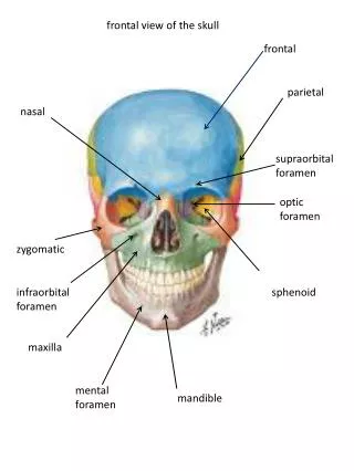
7. Embryology The critical period in nasal development is in first twelve weeks of fetal development
Abnormalities of development are believed to cause gliomas, dermoids, and encephaloceles
8. 3-4 weeks neural fold develops
Closure occurs from the midline and extends cranially and caudally
9. Neural crest cells play a key role in facial development
As the neural groove closes neural crest cells migrate around the eyes to the frontonasal process
10. In most of the body neural crest cells are involved in ectodermal components
In the face the primary role is mesenchymal cells
Bone, cartilage, and muscles of the face are all derivatives
11. Nose develops from frontonasal process and two nasal placodes
Medial processes fuse
Nasal-maxillary groove becomes the nasolacrimal duct
12. Mesenchymal structures form in centers which fuse
Key spaces are the foramen cecum, fonticulus nasofrontalis, and the prenasal space
13. Dermoids Cyst or sinus
Most common congenital midline mass
1-3% of all dermoids
3-12% dermoids of head and neck
14. ectodermal and mesodermal
Midline nasal pit, fistula, or infected mass from glabella to columella
Sometimes single cutaneous tract with hair at opening
May secrete pus or sebaceous material
15. Complications Intermittent inflammation
Abscess
Osteomyelitis
Broaden nasal root
Meningitis
Cerebral abscess
16. CNS connection variably reported from 4-45%
Associated congenital anomalies 5-41%
17. Associated Abnormalities Aural atresia, mental retardation, spinal column abnormalities, hydrocephalus, hypertelorism, hemifacial microsomia, albinism, corpus callosum agenesis, cerebral atrophy, lumbar lipoma, dermal cyst of the frontal lobe, coronary artery anomaly, cleft lip and palate, tracheoesophageal fistula, cardiac, genital and cerebral anomalies
18. Development During development a projection of dura projects through the foramen cecum
If skin maintains attachment to underlying fibrous tissue, nasal capsule, or dura epithelial elements may be pulled into the prenasal space with or without dural connection
20. Gliomas Glial cells in a connective tissue matrix
Red or bluish lump
Glabella, nasomaxillary suture, intranasal
Firm, noncompressible
Do not enlarge with crying
Do not transilluminate
May have telangiectasias
21. Extranasal 60%
Intranasal 30%
Both 10%
Dural connection 35% intranasal, 9% extranasal
Overall 15% dural connection
CSF rhinorrhea, meningitis
22. Develop from extracranial rests of glial tissue
Abnormal closure of fonticulus nasofrontalis, possibly encephaloceles which have lost CSF connection
23. Encephaloceles Extracranial herniation of meninges and/or brain
Subarachnoid connection
Rare at 1:35,000 births
1:6000 live births in Southeast Asia and Russia
30-40% associated anomalies: microcephaly, hydrocephalus, microopthalmia, anopthalmia, agenesis of the corpus callosum, porencephaly, cortical atrophy, ventricular dilations
25. Bluish, soft, compressible, transilluminate, pulsatile
Enlarge with crying
Positive Furstenberg test (bilateral compression of internal jugular veins)
Originate medially in the nose
May have associated CSF leak
26. Divided into three categories:
Occipital 75%
Sincipital 15%
Basal 10%
Sincipital-dorsum of nose, orbits, forehead
Basal- intranasal mass, nasopharynx, posterior orbit
27. Sincipital encephaloceles Nasofrontal
Nasoethmoidal
Nasoorbital
28. Basal Encephaloceles Transethmoidal-through cribiform plate into middle meatus
Sphenoethmoidal-extends through cranial defect between posterior ethmoids and sphenoid to nasopharynx
Transsphenoidal-presents in nasopharynx
Sphenomaxillary- through superior and inferior orbital fissures to sphenomaxillary fossa
29. Development Dural projection through fonticulus nasofrontalis
Abnormal closure results in herniated meninges/brain
May be closely related to glioma
30. Evaluation Most often infants and children
Dermoids-fistula tract, hair, pus or sebum, midline
Gliomas- firm, noncompressible, does not transilluminate, telangiectasias
Encephaloceles- soft, compressible, bluish or red, enlarge with crying, positive Furstenburg test
31. Do not biopsy extra or intranasal mass in a child before imaging
Risk of meningitis or CSF leak
Get imaging before biopsy
32. Imaging CT and MRI most used
CT findings include: fluid filled cyst, soft tissue mass, intracranial mass, enlargement of foramen cecum, distortion of crista galli
CT findings suggestive of intracranial extension are enlarged foramen cecum and bifidity of crista galli
Pensler et al reported that these findings were only valuable if absent, (when present may be false positive)
34. MRI Better delineates soft tissue
Ability to visualize in the sagittal plane
Denoyelle 36 children with dermoids, 2 patients had CT suggestive of intracranial involvement not found at surgery.
Recommended CT followed by MRI to confirm intracranial connection
35. Bloom et al
10 patients with nasal dermoids
One false positive, one indeterminate CT
Recommends MRI as first-line test due to increase cost of multiple tests, delay in diagnosis, additional risk if anesthesia required
37. Surgical Treatment Complete excision
Perform early to avoid nasal distortion, bony atrophy, osteomyelitis, meningitis
Denoyelle et al reported a recurrence rate of 5.5% of dermoids-must excise entire tract
38. Dermoids Pollock reviewed surgical treatment:
Access to all midline cysts and ability to perform medial and lateral osteotomies
Exposure for repair of cribiform defects, permit control of CSF rhinorrhea
Allow reconstruction of nasal dorsum
Offer probability of a favorable scar
39. Transverse rhinotomy
Small to moderately sized lesions
Avoids vertical scar and splaying
40. Transverse Rhinotomy Fistulous opening excised with transverse fusiform incision
Tract cannulated with lacrimal probe
Second incision made over dermoid
41. Tripod-eversion rhinotomy Larger lesions, especially lower 2/3 of nose
Transverse columellar incision, transfixion incision carried laterally and between upper and lower lateral cartilages
42. Paraalar incisions will permit upward rotation of the nose
Tract opening-fusiform excision
Cannulate fistulous tract
Operating microscope may be used to improve visualization
43. Zig-Zag rhinotomy Large lesions
Underlying bone or cartilage damage
Known intracranial extension
Provides wide exposure
Scar improved over straight vertical rhinotomy
44. Incisions designed with limbs which extend vertically >40 degrees <90 degrees
Fusiform excision of fistulous tract opening
Scar improved over straight vertical rhinotomy, limbs less than 90 degrees to RSTL running horizontally across the nose
45. Rohrich recommends open rhinoplasty for the following reasons:
Improved aesthetic results
Ease of exposure
Wide exposure of entire nasal dorsum
More control over osteotomy
Better visualization of the cribiform plate
46. Weiss et al described two cases of endoscopic dermoid excision in lesions with little or no skin involvement
47. Inferior portion removed via bilateral intercartilaginous and membranous septum incision
Aufricht retractor in place 0 and 30 telescope sinus tract followed to defect
48. Lateral rhinotomy may be used for intranasal gliomas or combine intra-extranasal
Several authors have reported isolated cases of endoscopic excision of small gliomas without evidence of intracranial extension.
49. Encephaloceles Will require combined approach with neurosurgery
Frontal craniotomy is performed, intracranial mass excised, bone-dura defect is repaired
Extracranial mass is then removed
Turgut et al reported mortality of 46% when there is brain tissue in the encephalocele
50. Key Points Midline nasal masses are rare but must be remembered in the differential
Don�t biopsy without imaging
Furstenberg�s test