Download
1 / 20
470 likes | 4.68k Views
Implantation. Begins 6-7 days after ovulation when the trophoblasts adhere to the endometrium The trophoblasts proliferate and form two distinct layers -Cytotrophoblast – cells of the inner layer that retain their cell boundaries

E N D
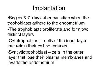
Implantation Begins 6-7 days after ovulation when the trophoblasts adhere to the endometrium The trophoblasts proliferate and form two distinct layers -Cytotrophoblast – cells of the inner layer that retain their cell boundaries -Syncytiotrophoblast – cells in the outer layer that lose their plasma membranes and invade the endometrium
Blastocyst during implantation 1. Blood vessel2. Endometrial stroma3. Syncytiotrophoblast4. Cytotrophoblast5. Surface epithelium6. Epiblast7. Aminotic cavity8. Hypoblast
types • 2 types: Interstitial implantation - occurs when embryo is embedded into uterine mucosa • Lies within the thickness of endometrium • Normal implantation. Intramural implantation- embryo sinks beneath the endometrium to reach the myometrium, also normal
Implantation site 1. Endometrium2. Uterine wall3. Implanted blastocyst4. Uterine tube
Implantation contd Enabled by estrogen and progesterone secretion The blastocyts embeds itself within the nutrient-rich uterine wall Takes approximately one week Human chrorionic gonadotropin (hCG) is secreted by the syncytiotrophoblast cells -Triggers the corpus luteum to secrete progesterone and estrogen -Prevents the uterine wall from shedding -Detectable by approximately one week following fertilization -Highest levels of hCG are around the second month, but declines as the placenta becomes functional
Implantation contd • Viability of the corpus luteum is maintained by human chorionic gonadotropin (hCG) secreted by the trophoblasts • hCG prompts the corpus luteum to continue to secrete progesterone and estrogen • Chorion – developed from trophoblasts after implantation, continues this hormonal stimulus • Between the second and third month, the placenta: • Assumes the role of progesterone and estrogen production • Is providing nutrients and removing wastes
Implantation involves the following process; (1) dissolution of the zona pellucida (2) orientation and adhesion of the blastocyst onto the endometrium (3) trophoblastic penetration into the endometrium (4) migration of the blastocyst into the endometrium (5) spread and proliferation of the trophoblast, which envelops and specifically disrupts and invades the maternal tissues. The embryo is drawn into a tight association with the uterine epithelium; uterine fluid is withdrawn by progesterone-sensitive pinopods, short-range forces enable embryos to adhere to epithelia. The pentasaccharide lacto-N-fucopentose-1 on the epithelium and its receptor on the trophoblast could be specifically involved in adhesion. contd
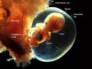
Summary of events of 2nd week • The blastocyst initially attached to the uterine endometrium (adplantation), • syncitiotrophoblasts then secrete enzymes that digest extracellular matrix, allowing the blastocyst to sink into the uterine wall, • majority of growth occurs in the trophoblastic shell. • The inner cell mass divides initially into 2 layers; epiblast and hypoblast (bilaminar embryo). • Hypoblast cells migrate around the original blastoceol cavity forming the primary yolk sac. • A second cavity (amniotic) forms between the inner cell mass and the cytotrophoblast shell; • this cavity is lined by epiblast cells.
Decidua • Part of the endometrium where the embryo is attached. • Decidual reaction- accumulation of glycogen and lipids by CT around the implantation site, and assumption of polyhedral shape. • cellular changes, together with the vascular and glandular alterations in the endometrium, are called the decidual reaction
1. Decidua capsularis2. Uterine wall3. Uterine cavity4. Placenta5. Decidua parietalis6. Decidua basalis7. Chorion leave8. Embryo9. Connecting stalk10. Yolk sac11. Chorion frondosum12. Amnion13. Chorionic cavity14. Amniotic cavity
decidua • Three different regions of the decidua are identified according to the implantation site. • The decidua basalis is the portion of the endometrium that underlies the implantation site. • The decidua basalis forms a compact layer, called the decidual (basal) plate. • The decidua capsularis is a thin portion of the endometrium that overlies the conceptus. • The decidua parietalis (vera) includes the remaining endometrium of the uterus and the cervix.