Download

1 / 46

E N D
Introduction to Endocrinology Physiology of the endocrine system by Dr Ehsan Saboory Professor of physiology Department of physiology, Faculty of medicine, Urmia University of Medical Sciences
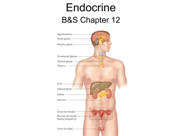
Endocrine System: Overview • Endocrine system – the body’s second great controlling system which influences metabolic activities of cells by means of hormones • Endocrine glands – pituitary, thyroid, parathyroid, adrenal, pineal, and thymus glands • The pancreas and gonads produce both hormones and exocrine products • The hypothalamus has both neural functions and releases hormones • Other tissues and organs that produce hormones – adipose cells, pockets of cells in the walls of the small intestine, stomach, kidneys, and heart
Hormones • Hormones – chemical substances secreted by cells into the extracellular fluids • Regulate the metabolic function of other cells • Have lag times ranging from seconds to hours • Tend to have prolonged effects • Are classified as amino acid-based hormones, or steroids • Eicosanoids – biologically active lipids with local hormone–like activity
Types of Hormones • Amino acid–based – most hormones belong to this class, including: • Amines, thyroxine, peptide, and protein hormones • Steroids – gonadal and adrenocortical hormones • Eicosanoids – leukotrienes and prostaglandins
Hormone Action • Hormones alter cell activity by one of the following mechanisms: • Direct changes in cell membrane permeability • Second messengers involving: • Regulatory G proteins (amino acid–based hormones) • Direct gene activation involving steroid hormones • The precise response depends on the type of the target cell and its receptor
Mechanism of Hormone Action • Hormones produce one or more of the following cellular changes: • Alter plasma membrane permeability • Stimulate protein synthesis • Activate or deactivate enzyme systems • Induce secretory activity • Stimulate mitosis
Amino Acid–Based Hormone Action: cAMP • Hormone (first messenger) binds to its receptor, which then binds to a G protein • The G protein is then activated as it binds GTP, displacing GDP • Activated G protein activates the effector enzyme adenylate cyclase • Adenylate cyclase generates cyclic AMP (cAMP) (second messenger ) from ATP • cAMP activates protein kinases, which then cause cellular effects
An Increase in cAMP Leads to: Activation of PKA which may cause : • Activationor deactivation of numerous enzymes • CREB→CREB-P + transcription factor 1→add to CRE • Stimulation or inhibition of RNA polymerase • Transcription of genes • cAMP finally hydrolyzed by phosphodiestrase (PD) • Activity of PD is also modulated by hormones via a G protein( dual regulation) • Two hormones can function antagonistically of one stimulates AC and the other stimulates PD
Amino Acid–Based Hormone Action: cAMP Second Messenger Figure 15.1a
Amino Acid–Based Hormone Action: PIP–Calcium • H binds to the receptor and activates G protein • G protein binds and activates a PLC enzyme • PLC splits the PIP2 into DAG and IP3(both act as second messengers) • DAG activates protein kinase C; IP3 triggers release of Ca2+ stores (PKC is Ca2+ dependent) • Ca2+ (third messenger) alters cellular responses • Arachidonic acid is derived from hydrolysis of DAG serve as a substrate of prostaglandins (PG) • The PGs are also modulators of hormonal response
Amino Acid–Based Hormone Action: PIP–Calcium Figure 15.1b
Other types of Signal transduction (ST) • 2 other mechanisms of ST from surface R are known in which the transducers lie in the cytoplasmic tail of the R • In one of these, H+R→ autophosphorylation of R→ the R itself becomes a tyrosine kinase and P the tyrosine residues on intracellular protein. Tyrosine P initiates a cascade of serine and threonine P of enzymes • Causes multiple intracellular events (Metabolism, proliferation and differentiation), e.g. Insulin • In the second one H+R→ conformational changes in R which attracts and docks tyrosine kinases e.g. GH • Another second messenger is cGMP→ activates PKG
Steroid Hormones • Steroid hormones and thyroid hormone diffuse easily into their target cells • Once inside, they bind and activate a specific intracellular receptor • The hormone-receptor complex travels to the nucleus and binds a DNA-associated receptor • This interaction prompts DNA transcription to produce mRNA • The mRNA is translated into proteins, which bring about a cellular effect
Steroid Hormones Figure 15.2
Intracellular receptors (steroid) • These R are large oligomeric and usually phosphorylated • Related to cis-oncogens family and contain 3 domains : • Variable C-terminus binds the H and is unique to each R • middle domain contains a DNA site formed by 2 fingers • N-terminus domain is variable in length( transactivating) • Unoccupied R is inactive or blocked by a molecule • H binding displaces the blocking molecule and translocates into the nucleus, undergo dimerization, binds to a specific site on DNA (HRE) → activates the RNA polymerase • Negative regulatory elements also exist in DNA molecules • These are characteristic of adrenal steroid hormones
Intracellular receptors (thyroxin) • In another general model of activation which is characteristic of thyroid hormones and Vit D the unoccupied R is already attached to DNA binding sites and prevents gene transcription • H binds R and relieves the suppressive effect of the R • In a variant of this model, H binding causes dissociation of the two identical receptor monomers constituting the homodimer, formation of heterodimer which activates the gene transcription
Hormone–Target Cell Specificity • Hormones circulate to all tissues but only activate cells referred to as target cells • Target cells must have specific receptors to which the hormone binds • These receptors may be intracellular or located on the plasma membrane • Examples of hormone activity • ACTH receptors are only found on certain cells of the adrenal cortex • Thyroxin receptors are found on nearly all cells of the body
Target Cell Activation • Target cell activation depends upon three factors • Blood levels of the hormone • Relative number of receptors on the target cell • The affinity of those receptors for the hormone • Up-regulation – target cells form more receptors in response to the hormone • Down-regulation – target cells lose receptors in response to the hormone
Receptor Kinetics • H+R =HR , K= HR/[H][R] , [HR]/[H]= K × [R] • H= free hormone in solution R=unoccupied receptor HR=bound hormone =occupied receptor R0= initial receptor capacity = [R] + [HR] K = affinity constant
Schatchard plot for HR kinetic A linear plot results when the H reacts with a single R class and no cooperativity is present. The negative of the K equals the slope of the line. The R number,R0, equals the intercept with the X axis.
Schatchard plot for HR kinetic An exponential plot results when the H occupancy of one R molecule alters the local affinity of a second nearby molecule for the H. This phenomenon called negative cooperativity.
A, The general shape of a H dose-response curve. Sensitivity is expressed as the concentration of the H that produces half-max response. B, Alterations in dose-response curve results from changes in max responsiveness, sensitivity, or both.
Hormone Concentrations in the Blood • Concentrations of circulating hormone reflect: • Rate of release • Speed of inactivation and removal from the body • Hormones are removed from the blood by: • Degrading enzymes • The kidneys • Liver enzyme systems
Control of Hormonal Secretion • Feedback control • Positive feedback: • Stimulus detected, H released, H reaches target cell, H binds R, Effect produced, Feedback to endocrine structure, More H released • Negative feedback: • Stimulus detected, H released, reaches target cell, binds R, Effect produced, Feedback to endocrine structure, Hormone release shut down • Direct nerve control • Autonomic nervous system • Inhibiting hormones or Releasing hormones • Chronotropic control
Feedback Neural Chronal
Control of Hormone Synthesis and Release • Blood levels of hormones: • Are controlled by negative feedback systems • by positive feedback systems (seldom) • Vary only within a narrow desirable range • Hormones are synthesized and released in response to: • Humoral stimuli (substrate or mineral- hormone) • Neural stimuli • Hormonal stimuli (hormone- hormone)
Humoral Stimuli • Humoral stimuli – secretion of hormones in direct response to changing blood levels of ions and nutrients • Example: Declining blood Ca2+ concentration stimulates the parathyroid glands to secrete PTH (parathyroid hormone) • PTH causes Ca2+ concentrations to rise and the stimulus is removed Figure 17.3a
Neural Stimuli • Neural stimuli – nerve fibers stimulate hormone release • Preganglionic sympathetic nervous system (SNS) fibers stimulate the adrenal medulla to secrete catecholamines Figure 15.3b
Hormonal Stimuli • Hormonal stimuli – release of hormones in response to hormones produced by other endocrine organs • The hypothalamic hormones stimulate the anterior pituitary • In turn, pituitary hormones stimulate targets to secrete still more hormones Figure 15.3c
Nervous System Modulation • The nervous system modifies the stimulation of endocrine glands and their negative feedback mechanisms • The nervous system can override normal endocrine controls For example, control of blood glucose levels • Normally the endocrine system maintains blood glucose • Under stress, the body needs more glucose • The hypothalamus and the sympathetic nervous system are activated to supply ample glucose
Chronal control (the circadian rhythms=CR) The origin of CR in H secretion, behavioral and metabolic activity. A clock with a 24-25h cycle is located in the SCN, this free running clock is entrained by environmental light signals to the external 24h day. It has bidirectional relationship with the sleep-wake cycle,too.
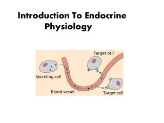
Type of cell to cell signaling • Endocrine: hormone enters the blood stream • Neurocrine (neuroendocrine):also enters the blood • Paracrine:through Int. fluid or GJ to another cell type • Autocrine: through Int. fluid or gap junction and act on neighboring identical cells or back to the cell of origin • Juxtacrine ?
Endocrine → Neurocrine → Paracrine → Autocrine →
Types of hormone synthesis • Protein or peptide hormone synthesis • Aminoacid based hormone synthesis • Steroid hormone synthesis • Eicosanoids synthesis
Hormone release • Release of protein and catecholamine hormones • Release of Thyroid and steroid hormones • Other forms of hormone release • Two adjacent cell types in a single gland may interact so that Hormone A (androgen) from cell A is modified in cell B to produce Hormone B (estrogens) • Modification of a precursor molecule of low activity to one of higher activity by successive steps (calcitriol) • Peptide Hormone can be produced in the circulation itself from a protein precursor (angiotensin)
Hormone Transport • Free • Bound to plasma proteins
Hormone Disposal • Irreversible removal of H is a result of: • Target cell uptake • Metabolic degradation • Urinary excretion • Biliary excretion • The sum of all removal processes is expressed as Metabolic Clearance Rate (MCR) • MCR= mg/min removed /mg/ml of plasma • K= MCR / volume of distribution • K is the fractional turnover rate • The plasma half-life is inversely related to K
Hormone measurement • The most common and useful method for measuring hormones is RIA (radioimmunoassay) • Estimate of hormone secretion rate • (V con – A con) × blood flow • This is useful in animal modal but not in human • Production Rate (PR) is suitable in human • PR is the total amount of the H entering the circulation per unit time • PR = Plasma con × MCR • Plasma levels is a valid index of Hormone PR
Location of the Major Endocrine Glands • The major endocrine glands include: • Pineal gland, hypothalamus, and pituitary • Thyroid, parathyroid, and thymus • Adrenal glands and pancreas • Gonads – male testes and female ovaries Figure 15.4