Download
1 / 46
810 likes | 2.29k Views
29.04.2015. HISTOLOGY OF GASTROINTESTINAL TRACT. Dr. Archana Rani Associate Professor Department of Anatomy KGMU UP, Lucknow. Contents. Oesophagus Stomach Small Intestine Large Intestine. Histology of the Digestive System. Basic Histological Layers: Mucosa: a. Epithelium

E N D
29.04.2015 HISTOLOGY OF GASTROINTESTINAL TRACT Dr. Archana Rani Associate Professor Department of Anatomy KGMU UP, Lucknow
Contents • Oesophagus • Stomach • Small Intestine • Large Intestine
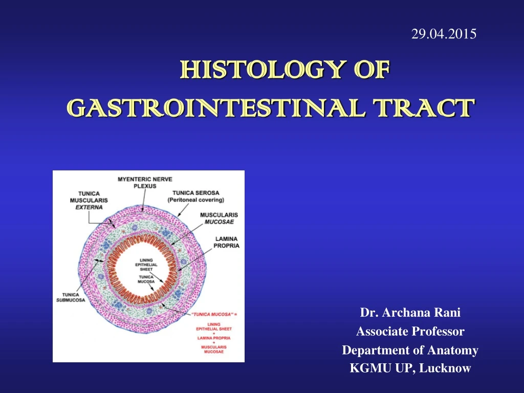
Histology of the Digestive System Basic Histological Layers: • Mucosa: a. Epithelium b. Lamina Propria c. Muscularis Mucosae • Submucosa: Submucosal plexus “Plexus of Meissner” • Muscularis: Myenteric plexus “Plexus of Auerbach • Serosa
Oesophagus • Mucosa: Stratified squamous non - keratinized epithelium • Submucosa: contains Meissner’s plexus and oesophageal glands • Muscularis externa: Upper one-third: skeletal fibres Middle one-third: mixed fibres Lower one-third: smooth fibres • Adventitia: loose areolar connective tissue
Stomach • Mucosa:simple columnar epithelium and presence of gastric pits. • Stomach is divided into three histological regions on the basis of nature of glands: • Cardiac region • Fundic region (fundus & body) • Pyloric region
Stomach (Cardiac Region) • Mucosa: simple columnar with oval nuclei, mucous secreting cardiac glands in lamina propria. • Submucosa: connective tissue. • Muscle layer: inner circular, outer longitudinal. • Serosa: simple squamous epithelium.
Stomach (Fundic Region) • Mucosa: simple columnar with oval nuclei, presence of gastric glands in lamina propria.
Stomach (Fundic Region) Cells of fundic region: • Mucous neck cells • Parietal (oxyntic) cells • Chief (peptic/zymogen) cells • Enteroendocrine cells • Undifferentiated cells
Stomach (Fundic Region) • Submucosa: contains blood vessels, lymphatics and Meissner’s plexus. • Muscularis Externa: an inner oblique (absent in pylorus), middle circular and outer longitudinal layer. • Serosa:consist of surface layer of flattened mesothelial cells resting on a thin layer of loose connective tissue with blood vessels and lymphatics.
Stomach (Pyloric Region) • Mucosa: pyloric glands in lamina propria & deeper gastric pits extending half the thickness of mucosa. • Muscularis Externa: inner circular (thickened to form pyloric sphincter) and outer longitudinal layer. • Submucosa & Serosa: same as in fundic part.
Small Intestine It is divided into duodenum, jejunum and ileum. • Mucosa: characteristic features- • Plicae circularis (valves of Kerkring) • Villi & Microvilli • Goblet cells (few) • Crypts of Lieberkuhn (intestinal glands) • Glands are lined by columnar cells, goblet cells, Paneth cells & enteroendocrine cells
Small Intestine • Submucosa: contains blood vessels, lymphatics and Meissner’s plexus. • Muscularis externa: Outer longitudinal and inner circular layers of smooth muscle. • Serosa/Adventitia
Duodenum Presence of Brunner’s glands in submucosa
Jejunum • Villi are tongue shaped. • Absence of Brunner’s glands.
Ileum • Presence of lymphoid aggregations in lamina propria known as Peyer’s patches. • Villi are short & finger like.
Large Intestine • It consists of: appendix, colon, rectum and anal canal. • Mucosa: Absence of Plicae circulares and villi Presence of Microvilli Presence of Crypts of Lieberkuhn Presence of Goblet cells in large number • Submucosa • Muscularis externa: Inner circular layer - thin compared to small intestine. Outer longitudinal layer- forms Taenia coli. • Adventitia: Appendices epiploicae (peritoneum forms pouch like processes filled with fat)
Vermiform Appendix • A small blind-ending diverticulum. • Largeaccumulations of lymphoid tissue in lamina propria which may extend into submucosa. • Intestinal villi are usually absent. • Crypts are poorly formed. • Muscularis externa is thin. • Absence of taenia coli.
Rectum • Intestinal glands are straight, like test tubes. • A continuous coat of longitudinal muscle is present. • Absence of taenia. • Absence of appendices epiploicae.
Anal Canal • Epithelium: upper part-simple columnar, middle part-stratified squamous non-keratinized, lower part-covered by true skin. • Mucosa has characteristic longitudinal folds-Anal columns. • Small mucosal folds between the anal columns -Pectinate line. • Crypts disappear below this line. • Muscularis externa-circular muscle forms involuntary internal anal sphincter.
References 1. diFiore’s Atlas of Histology with functional Correlations, 12th Edition. 2. Textbook of Human Histology. Inderbir Singh, 1st Edition. 3. Textbook of Histology. GP Pal, 3rd Edition.
MCQ Q1. Stratified squamous non-keratinized epithelium is a feature of: • Oesophagus • Stomach • Appendix • Rectum
MCQ Q2. Deep gastric pits is a feature of: • Oesophagus • Cardiac part of stomach • Fundic part of stomach • Pyloric part of stomach
MCQ Q3. Plicacircularis is a feature of: • Oesophagus • Stomach • Small intestine • Large intestine
MCQ Q4. Taenia coli is present in: • Oesophagus • Stomach • Small intestine • Large intestine
MCQ Q5. Abundant lymphoid tissue in lamina propria is a feature of: • Oesophagus • Stomach • Duodenum • Appendix