Download
1 / 32
640 likes | 2.11k Views
Histology of Respiratory Tract. Dr. Sama ul Haque Dr . Safaa. Objectives. Differentiate between the olfactory and nasal mucosa. Discuss the microscopic structure of larynx. Describe the microscopic structure of trachea and lungs.

E N D
Histology of Respiratory Tract Dr. SamaulHaque Dr. Safaa
Objectives • Differentiate between the olfactory and nasal mucosa. • Discuss the microscopic structure of larynx. • Describe the microscopic structure of trachea and lungs. • Differentiate between the terminal and respiratory bronchioles. • Explain alveolar cells.
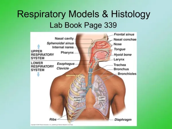
Respiratory System • This consists of the lungs and the air passages that lead to and form the lungs. • The air passages branch as they enter the lungs to finally form alveoli. • This system has 3 functions: 1.Air conduction; 2.Air filtration; 3.Gas exchange (respiration).
Air passages consist of: • A conducting portion • A respiratory portion • Conducting portion • 1.Nasal cavities; 2.Nasopharynx and oropharynx; 3.Larynx; 4.Trachea; 5.Paired primary bronchi. Bronchi within the lungs branch extensively to form bronchioles. The terminal bronchioles form the last part of the conducting system.
The respiratory portion is the part of the tract where gaseous exchange takes place and includes: 1.Respiratory bronchioles; 2.Alveolar ducts; 3.Alveolar sacs; 4.Alveoli.
Respiratory Epithelium • The typical respiratory epithelium consists of 5 cell types: 1.Ciliated cells; 2.Goblet cells; 3.Brush cells (have short blunt microvilli); 4.Small granule cells (contain secretory granules); 5.Basal cells (stem cells).
Nasal Cavity • Keratinized stratified squamous epithelium • Non Keratinized stratified squamous epithelium • Pseudostratified ciliated columnar epithelium with goblet cells • Olfactory Epithelium
Nasal Cavity • Nasal Cavity formed of: • 1.Vestibule. 2.Respiratory segment. 3.Olfactory segment • Vestibule: Keratinized stratified squamous epithelium Non Keratinized stratified squamous Contains vibrissae (fine hair), sebaceous glands and sweat glands
Nasal Cavity • Respiratory segment - Respiratory Epithelium - Lamina Propria • Olfactory segment - Olfactory Epithelium: i. Olfactory cells (bipolar neurons) ii. Basal cells iii. Supportive cells - Lamina Propria
Lamina Propria • Mucous Glands • Serous Glands • Venous Sinuses • Mucoperiosteum or Mucoperichondrium • Secretions of nasal mucosa - Bactericides - Lysozymes
Nasopharynx Mucosa • Respiratory Epithelium • Stratified Squamous Epithelium Lamina Propria • Elastic Tissue • Mucus Gland
Larynx • Mucosa False vocal cords: Pseudostratified ciliated columnar epithelium True vocal cords: Non Keratinized stratified squamous epithelium • Intrinsic Muscles • Cartilages - Larger Laryngeal Cartilages (Hyaline)
Epiglottis • Mucosa Anteriorly (Lingual mucosa): Non Keratinized stratified squamous epithelium Posteriorly: (Laryngeal mucosa): Pseudostratified ciliated columnar epithelium • Elastic Cartilage
Trachea • Mucosa • Pseudostratified ciliated columnar epithelium • Lamina propia • Sub mucosa • Connective Tissue • Seromucousbronchial Glands • Hyaline Cartilage • Adventitia
Bronchi • Mucosa; • Pseudostratified ciliated columnar epithelium • Lamina propia • Muscularis; • Submucosa; • Connective Tissue • SeromucousGlands • Cartilage layer; • Adventitia. Connective Tissue • At 1 mm diameter, the cartilage plates disappear and it becomes a bronchiole.
Respiratory Bronchioles Alveolar Ducts And Alveoli
Respiratory Bronchiole Simple cuboidal epithelium
Alveolar Cells • Alveolar Epithelial Cells (Type I Pneumocyte) • Type II Pneumocyte • Interalveolar Pores • Alveolar Macrophages (dust cells) • Alveolar Surfactant
Respiratory Membrane • It is the area where gas exchange occurs. It is formed of: • Alveolar Epithelium. • Fused basement membranes of the alveolar epithelium and the capillary endothelium. • Capillary Endothelium.