Download
1 / 20
210 likes | 396 Views
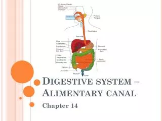
Digestive system – Alimentary canal . Chapter 14. Alimentary canal. Aka gastrointestinal (GI) tract Continuous tube open at both ends About 9m long in a cadaver; when living shorter due to muscle tone Food is technically outside the body. Mouth (oral cavity). Mastication (chewing) occurs

E N D
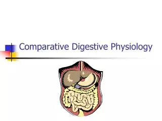
Digestive system – Alimentary canal Chapter 14
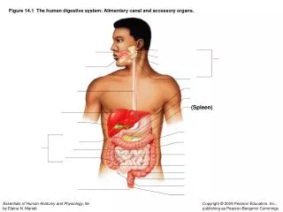
Alimentary canal • Aka gastrointestinal (GI) tract • Continuous tube open at both ends • About 9m long in a cadaver; when living shorter due to muscle tone • Food is technically outside the body
Mouth (oral cavity) • Mastication (chewing) occurs as saliva mixes w/ food • Parts • Lips • Cheeks –walls • Hard palate – anterior roof • Soft palate – posterior roof • Uvula – projection of soft palate • Vestibule – btw lips/cheeks and teeth/gums • Oral cavity proper – area w/in teeth • Tongue • Attaches to 2 bones – hyoid, styloid process • Lingual frenulum – secures to floor • Palatine tonsils – posterior – sides • Lingual tonsils – on base of tongue
Pharynx • Undergoes peristalsis • Oropharynx – posterior to oral cavity • Laryngopharynx – joins esophogus
Esophagus • Joins pharynx and stomach • About 25cm long • 4 tissue layers (tunics) – esophagus to large intestine • Mucosa – inner • Lines lumen • Surface epithelium w/ connective lamina propria and smooth muscle • Submucosa – contains blood and lymphatic vessels, nerve endings
Esophagus cont. • Muscularisextrna • Inner circular layer and outer longitudinal layer of smooth muscle • Serosa – outermost – visceral peritoneum • Connected to parietal peritoneum by mesentery • Walls contain submucosal nerve plexus and myenteric nerve plexus • Regulates mobility and secretory activity of GI tract organs
Stomach • On left side of cavity • Regions • Cardiac region – around cardioesophogeal sphincter • Where food enters • Fundus – lateral to cardiac region • Body • Pyloric antrum – most activity here • Pylorus – terminal – around pyloric sphincter valve
Stomach cont. • Length about 25cm • Diameter varies • May hold about 4L of food • Forms rugae (folds) when empty • Greater curvature – convex lateral surface • Lesser curvature – concave medial surface • Joined to liver by lesser omentum (peritoneum) • Greater omentum – hangs down to insulate, cushion, and protect organs • Contains an extra muscle layer – an oblique layer • Allows churning and mixing
Stomach cont. • Lining has gastric pits w/ gastric glands that secrete gastric juices • Chief cells – protein enzymes – pepsinogens • Parietal cells – hydrochloric acid • Mucous neck cells – produce protective mucous • Enteroendocrine cells – gastrin • Chyme – processed food that enters intestine
Small intestine • Average length 2.5-7m • Extends from the pyloric sphincter to the ileocecal valve • 3 divisions • Duodenum – “12 finger lengths long” • Bile duct and pancreatic duct join to form the hepatopancreaticampulla and enter duodenum via the duodenal papilla • Allows pancreatic enzymes and bile to enter and complete digestion • Jejunum – “empty” • Ileum – “twisted intestine”
Small intestine cont. • Pyloric sphincter – controls movement of food into intestine • Wall modifications • Microvilli – aka brush border • Tiny projections to increase surface area • Villi • Fingerlike projections • Contain capillaries and a lacteal (modified lymphatic capillary) – absorbed nutrients move into capillaries
Small intestine cont. • Circular folds – aka plicacirculares • Deep folds in mucosa and submucosa layers – increase surface area • Peyer’s patches – collections of lymphatic tissue – increase toward end of small intestine to keep bacteria out of blood
Large intestine • About 1.5m long • Extends from ileocecal valve to the anus • Absorbs water from indigestible food and gets rid of wastes • Subdivisions • Cecum – sac-like • Appendix – hangs from cecum • Appendicitis – build up of bacteria
Large intestine cont. • Colon • Ascending colon – up right side of body – turns at right colic (hepatic) flexure • Transverse colon – moves across body – turns at left colic (splenic) flexure • Descending colon – moves down left side • Sigmoid colon – enters pelvis • Rectum • Anal canal – opens to exterior at anus • Voluntary sphincter (external anal sphincter) – skeletal muscle • Involuntary sphincter – smooth muscle
Large intestine cont. • Mucosa contains goblet cells – produce an alkaline mucus for lubrication • Haustra – puckering of wall due to muscle tone of teniae coli (longitudinal muscle)
Accessory organs – salivary glands • Empty into mouth • Parotid glands – anterior to ears – mumps • Submandibular glands – floor of mouth • Sublingual glands – floor of mouth • All produce saliva – mixture • Mucus – moistens and binds food into a bolus • Serous fluid – contains salivary amylase – begins starch digestion
Accessory organs - teeth • 1st set – deciduous teeth – baby teeth; milk teeth • Should have full set by 2 years • Begin to lose btw 6-12 years old • 2nd set – permanent teeth – 32 • Should have all but third molars (wisdom teeth) by end of adolescence • Wisdom teeth emerge btw 17-25 • Types – pairs listed for top ½ of mouth • Incisors (2 pr) – cutting • Canines (1pr)– tearing/piercing • Premolars (2 pr) and molars (3 pr) – grinding
Teeth cont. • Parts • Crown – exposed portion • Above gingiva (gum) • Covered by enamel • Dentin – bulk of tooth – under enamel • Pulp cavity – contains blood vessels and nerves • Neck – connects crown to root • Root – embedded in jawbone • Cementum – covers outer surface and attaches to periodontal membrane (holds tooth in place) • Root canal – where pulp cavity enters root
Accessory organs • Pancreas – produces digestion enzymes • Also produces insulin and glucagen • Liver – largest gland in body – 4 lobes • Lies over stomach • Hangs from diaphragm and abdominal wall by falciform ligament • Produces bile – leaves via the common hepatic duct • Composed of bile salts and phospholipids which aid digestion • Salts emulsify fats by breaking large globules into smaller ones
Accessory organ - gallbladder • Small, green sac on the inferior surface of the liver • Connected to liver via the cystic duct • Stores bile until needed • Gallstones – form when bile is stored too long or too much water is removed cholesterol crystallizes