Download

1 / 29
400 likes | 1.29k Views
CTVT. -- Chapter 32, pg. 1093-1148. VTDRG. --Pg. 497-520. Veterinary Dentistry. Ethical and Legal Aspects. The level of dental care that a veterinary technician may provide varies from state to state.

E N D
CTVT -- Chapter 32, pg. 1093-1148 VTDRG --Pg. 497-520 Veterinary Dentistry
Ethical and Legal Aspects The level of dental care that a veterinary technician may provide varies from state to state. The American Veterinary Dental College (AVDC) considers it appropriate for the veterinarian to delegate maintenance dental care and certain dental tasks to veterinary technicians.
Dental Tasks for Veterinary Technicians Oral examination and charting Taking and developing dental radiographs Dental prophylaxis Client education Taking impressions and making models Performing nonsurgical, subgingival root planing *Procedures performed by veterinary technicians must not result in alterations in the shape, structure, or positional location of teeth in the dental arch.
Veterinary Dental Organizations • Opportunities for advanced training in dentistry: • NAVTA responsible for governing and overseeing Veterinary Technician Specialists (VTS). • Anesthesia, emergency and critical care, internal medicine, dentistry, and behavior • Academy of Veterinary Dental Technicians (AVDT) credentials RVTs as specialists in dentistry • Requires 3000 hours experience then…secure a mentor, maintain case logs, write case reports, pass a specialty exam, and attend CE courses. • See www.avdt.us for further info.
Veterinary Dental Organizations • American Veterinary Dental Society (AVDS) • www.avds-online.org • American Society for Veterinary Dental Technicians (ASVDT) • www.asvdt.org
Tooth Classification • Brachyodont teeth: small crown with long, well-developed roots; teeth do not continue to grow or erupt throughout animal’s life. • Ex: humans, carnivores, pigs • Hypsodont teeth: long crown height and continue to erupt for most the animal’s lifetime. • Ex: horses, rodents, and lagomorphs • Diphyodonts (most mammals) have two sets of teeth, deciduous (baby teeth) then permanent.
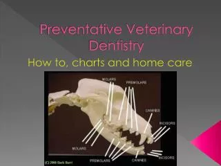
Oral Anatomy Upper teeth embedded in maxilla(upper arch). Lower teeth anchored in mandible (lower arch). Front, upper teeth are called incisors and are housed in the premaxilla(a.k.a. incisive bone). Maxilla houses the upper canine teeth and cheek teeth (molars and premolars). Palatine bone comprises most of the hard palate. Teeth of the mandible = jaw teeth What is the TMJ?
Dental Morphology • Incisors: in front of the mouth and are used for gnawing and grooming. • Canine Teeth: are distal to the incisors, very long and used for grasping and tearing. • Premolars and Molars: (a.k.a. cheek teeth), are used for shearing and grinding.
Dentition Mammals show great variety in dentition (numbers and types of teeth). Dental formulas are used to classify the normal dentitions of different animal species. Some species variation exists in normal eruption times of deciduous and permanent teeth.
Normal Dentition of Dogs and Cats • Puppies have 28 deciduous (primary / milk) teeth. • Eruption occurs at about 2 to 12 weeks • Dogs have 42 permanent teeth. • Eruption occurs about 3 to 7 months. • Kittens have 26 deciduous teeth. • Eruption occurs at about 2 to 6 weeks • Cats have 30 permanent teeth. • Eruption occurs about 3 to 6 months.
Normal Dentition of the Dog and Cat This represents one side of the mouth top/bottom 42 Note: Cats have one less lower premolar; dogs have one less upper molar
Covers the crown Very hard bone that makes up the majority of tooth. Above the gum line. Within the dentin, made up of nerves, blood vessels, and connective tissue Gingiva Covers the roots Below the gum line. Shock absorbing lining; attaches tooth to the bony socket. Bony socket
Tooth Morphology • Crown = part of the tooth that is visible in the mouth above the gumline. • Shiny enamel covers the crown of the tooth. • Hardest substance in the body. • Prevents tooth from being invaded by bacteria and acids. • If destroyed, will not regrow. • The root is the tooth structure below the gumline. • Root is covered by cementum. • The tip of the root is the apex of the tooth. This is where the nerve enters from the jaw into the tooth.
Tooth Morphology • Dentin is the substance that composes the bulk of the tooth. • Harder than bone but not as hard as enamel. • Lies under the enamel and cementum. • Produced by the pulp of the tooth which is composed of blood vessels, nerves, and connective tissue. • Pulp cavity is located within the central core of the tooth and contains pulp. • Pulp chamber is the portion of the pulp cavity located in the crown. • Root canal is the portion located beneath the gumline.
The Periodontum • The periodontiumis the area where the tooth meets the gum. It functions to attach the tooth to the jaw and provide support, resistant to normal functional forces. It includes: • Gingiva • Periodontal ligament • Cementum • Alveolar bone
The Periodontum • Gingiva= gum tissue; further divided into free gingiva, which lies closely against the tooth and attached gingiva, which is firmly attached to the underlying alveolar bone. • Gingival sulcusis the shallow groove between the tooth and the gingiva. • Formed by the rounded margin of the free gingiva. • Called a periodontal pocket when abnormal. • Mucogingival Line (MGL) is the junction line where the firm, attached gingiva connects to the loose alveolar (buccal) mucosa.
In Perspective…. MGJ- Mucoginival Junction AM- Alveolar Mucosa G- Gingiva (free gingiva)
The Peridontium • Periodontal ligament attaches cementum to the alveolar bone of the mandible or maxilla. • Cementumis a bone-like tissue that covers the root surface. It is stronger than bone but not as strong as enamel. • It is capable of repairing itself. • Alveolar bone forms the tooth socket. • Blood vessels and nerves run through the alveolar bone; most supply the periodontal ligament.
Dental Directional Terminology Rostral refers to a structure that is closer to the front of the head in comparison with another structure. Caudal describes a structure toward the back of the head when compared to another structure. Vestibular describes the tooth surface facing the lips or vestibule (buccal, labial).
Dental Directional Terminology Facial describes vestibular surface of teeth visible from the front (incisors). Lingual refers to the surface of the mandibular teeth adjacent to the tongue. Palatal refers to the surface of maxillary teeth adjacent to the palate
Dental Terminology Mesial refers to the portion of the tooth in line with the dental arcade that is closest to the most rostral portion of the midline of the dental arch. Distal refers to the portion of the tooth that is closest to the most caudal portion of the midline of the dental arch. Apical refers to a portion of the tooth closer to the apex, or tip of the root. Coronal refers to a structure within a location closer to the crown of the tooth in relation to another structure.
Dental Terminology Labial describes tooth surface facing lips. Occlusalrefers to the part of a tooth that meets with, or occludes with, the teeth of the opposite dental arcade. Buccalpertains to the inside of cheek or mouth. Interdental space refers to the space between each individual tooth.
A palatal view of the dog maxilla. The midline is marked with a line, and the mesial and distal tooth surfaces are marked with an M or D. The cingulum is visible on the maxillary incisors. Bump like located on the side of the incisors nearest the tongue Surface of tooth facing the next tooth forward Surface facing tooth behind
The mandibular first molar.The X’s indicate the cervical or gingival area of the teeth. Area between roots of multirooted teeth