Download
1 / 26
260 likes | 426 Views
A 57-year-old male from the Philippines presents with progressive fatigue and shortness of breath, leading to an admission for evaluation. His history includes gout, with recent symptoms of low-grade fevers and mild sore throat. Laboratory findings indicate severe anemia (Hgb 3.3) and elevated LDH, suggesting autoimmune hemolytic anemia (AIHA). Diagnostic workup reveals evidence of hemolysis with low haptoglobin and schistocytes. This case highlights the diagnostic approach and management of AIHA, underlining its classification and clinical implications.

E N D
Heme-Onc Subspeciality Rounds Dr.Chaudhry Dr.Vysetti
Case HPI:57 Yr old male from Philippines gets admitted for evaluation of progressively worsening fatigue and shortness of breath -2 wks. • Had a mild sore throat and headache about a week ago which resolved on its own. • Low grade fevers • ROS otherwise negative PMH: - Gout PSH: -Hand surgery involving the joints.
Case Meds: 1.Allopurinol 100mg po qdaily 2.Tylenol Prn. Family History: Non contributory Social History : Moved to US about a year ago. Currently unemployed. lives with his wife who is a nurse.
Case Vitals : Afeb P:102 B.p 80/50 98% on RA Exam : Gen: Positive for Icterus and pallor.No enlarged Lymphnodes RS:CTA CVS:S1S2 no murmers GI :Soft, Spleen palpable 4 cm below the left subcostal margin.liver not palpable.Otherwise Nontender ,BS + CNS : No focal deficits. Ext :No pedal odema. Labs on admission : CBC : Hgb 3.3 and Hematocrit of 13.5 (MCV – 98.2,MCH-41.9,MCHC-42.7,Bands 7%,Monocytes of 31%,Neutrophils -52%) CMP : Na :123,K:4.2 ,HCO3 17, Cl 93,BUN 18 and Creat 1.1,Total Bili 3.3,Alkphos 99,AST and ALT wnl. PT 18.7 & INR – 1.6
Hospital Course Diagnostic Work up : -LDH -1145, -Haptoglobin <6 , -Retic count : 13.6 - PBS:clumping of RBC’s at room temp which resolved upon warming the slides.No Schistocytes.
Hospital Course Follow up : -Daily CBC’s,Total Bilirubin,Serum haptoglobulin and LDH. -RBC count and platelets remain low but stable. -H&H – 6.0 -9.8 Most recent -6.4 -Haptoglobulin <6 until recently reported as 27 (ref 36-195) -LDH down to 464 from 1145.Recent uptrending 634.(132-268) -Total Bilirubin 6.8-3.5.(0.0-1.0)
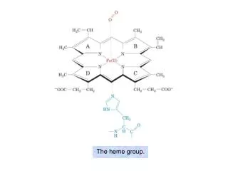
Autoimmune hemolytic Anemia -Autoimmune hemolytic anemia (AIHA) refers to a collection of disorders characterized by the presence of autoantibodies that bind to antigens on patient's own erythrocytes, leading to premature red cell destruction - Immune Disorder caused by antibodies directed against unmodified autologous red cells.
Pathophysiology IgG antibodies are relatively poor activators of complement and recognised more readily by the Fc Receptors on the phagocytic cells.So in turn destroyed by Phagocytes of the RES. IgM-sensitised RBC’s generally are associated with both intravascular and extravascular hemolysis.Intravascular because IgM unlike IgG more readily activate the complement pathway and produce intravascular hemolysis.Extravascular hemolysis occurs from RES being activated by the complement bound on the surface of the RBC. Spleen is the principal site of IgG associated extravascular hemolysis and Kupffer cells in the liver are the principal effectors of IgM associated extravascular hemolysis.
Classification of Immune hemolytic Anemia Warm AIHA Idiopathic Vs Secondary Cold AIHA CAS :Idiopathic Vs Secondary PCH : Idiopathic Vs Secondary Mixed AIHA Drug Induced immune hemolytic anemia Alloantibody-induced immune hemolytic anemia Secondary causes: Infections, Autoimmune , Lymphoproliferative disorders.
Diagnosis Clinical Presentation and lab evidence of hemolysis Serologic evidence of an Autoantibody
Clinical Presentation -History : Recent Meds and Infections & Systemic illness. -Physical findings : Anemia,Jaundice, Organomegaly. -Lab evaluation . Retic count-Increased . PBS –Spherocytes,Schistocytes,Cold Agglutinins. . Bone Marrow – r/o Malignancy.Erythroid Hyperplasia. . UA – If Intravascular hemolysis -Hemoglobinuria . Direct Globulin Test(Coomb’s) – identifies the presence of antibodies and/or complement on the surface of the erythrocyte. . Other – elevated total bilirubin,AST and decreased Haptoglobin . Immunohematologic studies-
Autoantibodies - Two major types, each with specific characteristics, are produced in AIHA: - IgG antibodies : Generally react with protein antigens on the RBC surface at 37 c and so called "warm agglutinins“. - IgM antibodies: Generally react with polysaccharide antigens on the RBC surface only at temperature 0-4 c and so called "cold agglutinins.”
Factors affecting the rate and location of erythrocyte destruction: Characteristics of the autoantibody - Antibody isotype (eg, IgG, IgM, IgA) - Antibody titer (high vs. low) - Thermal reactivity (optimal binding temperature) - Ability to fix complement . Characteristics of the erythrocyte antigen - Specificity of the antigen - Surface density of the antigen . Characteristics of the reticuloendothelial (RE) clearance - Preferential location of clearance (hepatic vs. splenic)
Direct Antiglobulin test - The RBCs of the patient are washed free of adherent proteins and reacted with antiserum or monoclonal antibodies prepared against the various immunoglobulins, particularly IgG and a fragment of the third component of complement, C3d. - Positive test –Presence of an autoantibody - Further the sample is tested seperately with reagents specific for anti IgG and Anti-C3d.
Warm Agglutinin Disease Incidence - 50%-70% cases Etiology : Idiopathic Vs Secondary( CLL,Hodgkins,NHL,Autoimmune ) Clinical course :Insidious onset with waxing and waning course Vrs Fulminant hemolysis. Diagnosis : Positive DAT and lab evidence of hemolysis. -Rx: - Steroids, If no response to steroids next line of Rx includes splenectomy and cytotoxic drugs. - Splenectomy has a response rate of 60-75%(removal of site of hemolysis ) • Cytotoxic drugs in people who failed steroids and/or splenectomy- response rate • of 40-60%(Cyclophosphamide and Azathioprine) - Other therapies :Rituximab (Anti –CD20 monoclonal AB),Plasmapheresis,?IVIG,Danazol - PRBC transfusions limited to life threatening hemolytic anemia.
Cold AIHA CAS PCH - More common • Middle-elderly • Prim Vs Sec (MC) • IgM • Max reactivity in the cold but reactive up to 30 c • Resistant to Rx - Less common • Children • Often follows a URI • IgG (DL) • Biphasic hemolysin demonstrated by incubation in cold followed by incubation at 37 c in presence of C. • Supportive Rx.
CAS Incidence : 16-32% Etiology : Primary Vs Secondary -Infections: Mycoplasma,EBV,Influenza,HIV,E.coli,listeria -Lymphoproliferative : CLL,Lymphomas,Waldenstrom’s. CF : -Mild chronic hemolytic anemia exacerbated by cold environmentepisodes of acute hemolysis common in winter months. -Clinical presentation corresponds with the immune response to an infectious agent. -Symptoms appear 2-3 wks after the infections begins(corresponding to Cold Agglutinin titre) and resolve 2-3 wks later.
CAS Diagnosis Treatment • Avoidance of cold exposure. • Therapy directed against secondary causes. • Immunosuppression with chlorambucil,cyclophosphamide. • Steroids are rarely helpful. • Splenectomy-? Since extravascular hemolysis (IgM mediated )occurs in Liver. • Plasmapheresis - Rituximab has shown success. - Typically Clumping of the RBC even before the antisera is added and dissolution of the clumping upon warming indicates the presence. - Positive DAT with Anti –C3 and often negative for IgG. - IgM can be Monoclonal and Polyclonal. - Antigens usually “ I “ on adult RBC -
PCH Incidence -Rare 2-10% Children MC affected Etiology: - Idiopathic Vrs Secondary - Infections Measles,Mumps,EBV,CMV,VZ,Adeno,Influenza,Mycoplasma,Hemophilus. Diagnosis : - DAT: positive with Anti-C3 but is generally negative for anti-IgG unless performed at colder temperatures. - Donath Landsteiner AB –Biphasic hemolysin - Antigen is P present on most of RBC Rx : - Generally self limited.No need for aggressive measures.
Drug Induced IHA • MC drugs : Alpha methyl dopa,PCN,2nd & 3rd Cephalosporins • Rarely Levodopa,Diclofenac. • CF:Variable MC subacute onset but rarely acute hemolysis. • Prognosis –Excellent. • Therapy –Stop drug and rarely empiric course of steroids.
Hospital Course Diagnostic Work up: - CMV IgG > 5.0 (ref <0.9) IgM Pending. - EBV cap AB,IgG- 1: 10,240 & IgM- Neg - Bone marrow Biopsy (Day 6)-Flow cyto:Small population of monoclonal B cell (1%).Final reports – negative for Acute leukemia,MDS,LPD,plasma cell dyscrasia. - Cold agglutinin titres 1:512 -Preliminary results : ID antibody as Anti-S -Mycoplasma IgG and IgM –ve. -Hep A,B and C – Non reactive - HIV – Non reactive - PNH :r/o by flow cytometry - SPEP : unremarkable .Low IgM - Donath Landsteiner Antibody negative r/o PCH.. - HIT panel negative (was ordered because of low platelet count).
Hospital Course • Admitted to ICU • Inetially transfused 4U of PRBC which brought his H/H to 6/13 - High dose IV steroids initiated on Day 3 x 3 d following by high dose oral prednisone (60mg po daily). • On day 2 pt had to be intubated because of ?TRALI ,was weaned off the ventilator over couple of days. • The pt continued to require daily transfusions.(received 17 U Prbc’s so far since admission). • Plasmapheresis initiated on Day 3 continued for total of 7 days. • Relative stabilisation of H&H after Plasmapheresis was initiated • Held for a couple of days and reinitiated because of persisting Hemolysis. • Danazol started on Day 3 (200mg po BID) • Cyclophosphamide (100mg po q daily) started on Day 10. • Rituximab (Received 3 doses so far
Hospital Course Follow up : -Daily CBC’s,Total Bilirubin,Serum haptoglobulin and LDH. -RBC count and platelets remain low but stable. -H&H – 6.0 -9.8 Most recent -8.0 and is stable -Haptoglobulin <6 until recently reported as 27 (ref 36-195) -LDH down to 464 from 1145.Recent uptrending 634.(132-268) -Total Bilirubin 6.8-3.5.(0.0-1.0)