Download
1 / 16
170 likes | 206 Views
Explore the revolutionary approach of nano-surgery for repairing individual axons, a critical step in treating nerve injuries. Discover the groundbreaking techniques and tools used in subcellular surgical procedures to restore neurological function. Learn about laser cutting and mechanical cutting methods, along with the innovative Nanoknife technology. This emerging field shows promise in addressing nerve damage on a microscale, with potential implications for neurological treatments. Despite challenges, research in this area continues to advance, shaping the future of axon repair methodologies.

E N D
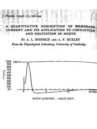
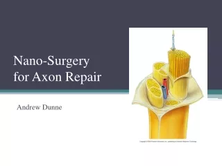
Nano-Surgeryfor Axon Repair Andrew Dunne
Background • Injury to the nervous system is a common occurrence after trauma. • Unlike some tissues in the body that exhibit self healing, nerve cells that are injured, particularly those in the brain and spinal cord, are incapable of regenerating circuits by themselves to restore neurological function • May lead to the loss of sensory/motor function.
Christopher and Dana Reeve Foundation • In the USA alone, close to 1.3 million individuals suffer from some form of spinal cord injury and live with permanent neurological dysfunction. • Impact on society increases greatly when considering PNS injuries. • Demonstrates the need for treatment of nerve injuries
Research Areas • Researchers explore whether micro/nanoscale tools and materials can be used to address this major challenge in neuromedicine. • Two Paths: • Stimulating axon regeneration • The development of new nanoscale tissue scaffold materials • Direct surgery on individual axons using micro devices as surgical tools.
Axon Surgery • This approach conceders nerve injury as a subcellular surgical problem in which micro and nano devices are used as instruments to repair of individual axons • Central Thesis: • Physical rejoining of two severed axon segments will restore axon electrical conduction • Beyond existing surgical technologies • Obstacles: • Technical inability to perform surgical manipulations at a subcellular scale on the axon • Progress has been made with testing of microdevices with nanoscale features to perform basic steps of axon repair
Basic steps • Removal of injured ends of axons and trimming back to healthy axon segments • Separated axon segments being physically brought together • Fusing the membranes of apposing axon segments to form a single structurally intact and functional axon.
Laser Cutting • Widely investigated method for biological cutting with micron or submicron scale precision • Nano-scissors • Have been used to cut or ablate whole cells and intracellular elements • Chromosomes, mitochondria and microtubules. • Use of low energy femtosecond laser pulses to create a high photon concentration in a small area without creating a lot of heat that might damage surrounding axons. • Dye labeling typically used to photosensitize the intended targets prior to ablation.
Experiment • University of Texas • Cut individual axons in nematode (1mm long) • Cut size 300-500nm in diameter • Cut a axon known to impair worms backward motion. • After surgery couldn’t move backwards. • 24 hours later grew back the severed axon proving the laser didn’t damage surrounding tissue.
Mechanical Cutting • Miniaturized mechanical cutting to cut individual axons • simpler, less costly, does not require dye photosensitization. • A Nanoknife • Produced using silicon microfabrication and microassembly techniques • Demonstrated for precise cutting of a variety of CNS and PNS axons both in vitro and in anesthetized animals in vivo
500µm Nanoknife • Two microfabricated silicon components • Micro fabricated frame and suspension • Blade in the shape of an elongated pyramid • Frame attached to a rod • Micromanipulator holds rod in place during use and delivers cutting stroke.
Nanoknife continued • 1mm2 frame and pair of serpentine flexures for multiaxial motion • Centered blade. • Cutting edge: 20nm radius of curvature and about the width of a synaptic cleft • Length: fabrication of one to hundreds of microns 20µm 100nm 200µm
Experiment • Extensively tested • Highly effective in severing axons (both myelinated and umyelinated) • Delivers cut without distorting adjacent segments and avoids mechanical shearing • Tested in vivo to address questions of whether miniaturized cutting instruments can be effective under real surgical conditions. • Strength • Demonstrated by making repeated cuts at axons from the sciatic nerve of a mouse. • Precision • Targeted removal of short axon segment • Mimics the first of the three proposed steps in axon surgical repair 25 µm
Single axon surgery on mouse Isolation of short segment using a Nanoknife Scale = 200 µm
Conclusion • Different techniques still being tested for the multiple steps in axon repair. • Cutting away • Moving of axon ends together • Fusion of the ends to make it whole • This is not a reality yet • Techniques from different stages must be brought together and researched even more before micro-technology based axon repair can be used in patients
Resources • Change, WC, Hawkes, E, Keller, CG, & Sretavan, DW. (2010, January 25). Axon repair: surgical application at a subcellular scale. John Wiley & Sons, Inc., Retrieved from http://www3.interscience.wiley.com/cgi-bin/fulltext/123261285/HTMLSTART doi: 10.1002/wnan.76 • http://en.wikipedia.org/wiki/Axon • http://www.christopherreeve.org • http://www.utexas.edu/news/2004/12/16/nr_engineering/