Download
1 / 28
300 likes | 556 Views
Learn about the anatomy, functions, and connections of the thalamus and limbic system. Understand the effects of lesions and the crucial role these structures play in brain function.

E N D
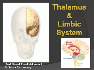
Thalamus & Limbic System Prof. Saeed Abuel Makarem & Dr.Sanaa Alshaarawy
Objectives By the end of the lecture, you should be able to: • Describe the anatomy and main functions of the thalamus. • Name and identify different nuclei of the thalamus. • Describe the main connections and functions of thalamic nuclei. • Name and identify different parts of the limbic system. • Describe main functions of the limbic system. • Describe the effects of lesions of the limbic system.
Thalamus • It is thelargest nuclear mass of the whole body. • It is the largest part of the diencephalon • It is formed of • two oval masses of grey matter. • It is the gateway to the cortex. • Together with the hypothalamus they form thelateral wall of the 3rd ventricle. THALAMUS Corpus callosum Midbrain PONS
It sends received information to the cerebral cortex from diverse brain regions. • Axons from every sensory system (except olfaction) synapse in the thalamus as the last relay site 'last pit stop' before the information reaches the cerebral cortex. • There are some thalamic nuclei that receive input from: • Cerebellar nuclei, • Basal ganglia- and • Limbic-related brain regions. Thalamus
Relations It has 4 surfaces & 2 ends. Surfaces Lateral:(L) Posterior limb of the internal capsule Medial: The 3rd ventricle It is connected to the thalamus of the opposite side by the interthalamic connexus, (adhesion) or Massa intermedia. Superior: (s) Lateral ventricle and fornix. Inferior: (I) Hypothalamus, anteriorly & Subthalamus posteriorly. S L I
Anterior end: Forms a projection, called the anterior tubercle. It lies justbehindthe interventricular foramen. Posterior end: Broad Forms a projection called Pulvinarwhich lies above the superior colliculus and the lateral & medial Geniculate bodies.
Internal Structure • White matter: • External medullary lamina: • Covers the lateral surface. • It consists of thalamocortical & corticothalamic fibers. • Internal medullary lamina: • Bundle of Y- shaped myelinated (afferent & efferent) fibers. • It divides the thalamus into: anterior , medial, lateralnuclear groups. • Each of these groups is subdivided into a number of named nuclei.
Lateral Nuclear Group It is divided into: Dorsal & Ventral tiers Dorsal tier: which contains: Lateral Dorsal (LD)& Lateral Posterior (LP) Pulvinar. Ventral tier, which contains : Ventral Anterior (VA) Ventral Lateral (VL) Ventral Intermediate (VI) Ventral Posterior (VP) (PLVNT, PMVNT) Medial & Lateral Geniculate nuclei.
Projection of thalamic nuclei Anterior Thalamic Nucleus • Afferent: Mammillary body. • Efferent: Cingulate gyrus, (limbic system) • ------------------------------- Medial Nucleus • Afferent: Hypothalamus. • Efferent: Frontal cortex.
Projection of thalamic nuclei Ventral Anterior Nucleus • Afferent: Globus pallidus body. • Efferent: Premotor cortex. • ------------------------------- Ventral Lateral Nucleus • Afferent: Dentate Nucleus • Efferent: primary motor cortex.
Projection of thalamic nuclei Ventral Posterior Lateral Nucleus • Afferent: Medial and spinal leminsci. • Efferent: Sensory cortex. • ------------------------------- Ventral Posterior Medial Nucleus • Afferent: Trigeminal Leminiscus • Efferent: Sensory cortex.
Projection of thalamic nuclei • Medial geniculate body : • Afferent : lateral lemniscus. • Efferent : auditory cortex. • Lateral geniculate body : • Afferent : optic tract. • Efferent : visual cortex
Frontal Primary motor cortex Pre-motor cortex Sensory cortex Cingulate gyrus
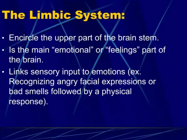
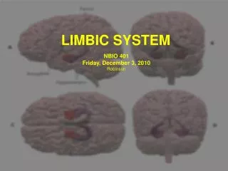
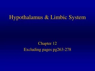
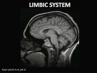
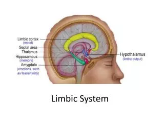
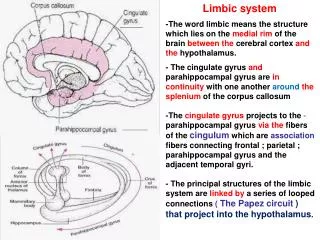
LIMBIC SYSTEM • The term "limbic" is from the Latin word Limbus, for "border" or "edge". • It separates the medial surface of the cerebral cortex from the diencephalon • It consists of a number of cortical&subcorticalstructures with looped connections that all project to the hypothalamus.
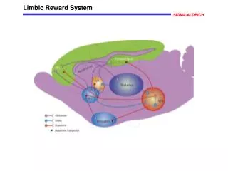
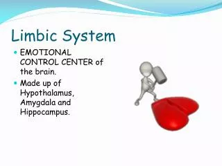
WHAT IS THE FUNCTION OF THE LIMBIC SYSTEM? Pleasure sensation It control a variety of functions including: • Emotions, • Emotional responses • Behaviour & Mood (happy, cry, laugh, sad, afraid, aggression, depression) • Motivation. • Memory. • Visceral & Motor responses involved in (sex, pleasure, hunger, and reproduction). • Olfaction. OLFACTION MEMORY
The limbic system is composed offour main structures: • 1. Limbic cortex • 2. Amygdala. • 3. Hippocampus, & • 4. Septal area. • These structures form connections between the limbic system and the hypothalamus, thalamus and cerebral cortex. • The hippocampus is important in memory and learning, while the limbic system itself is important in the control of the emotional responses. The limbic system is a set of brain structures including
CORTICAL STRUCTURES • Limbic lobe. • Hippocampal formation. • Septal areas. • Prefrontal area.
LIMBIC LOBE • C-shaped ring of grey matter on the medial side of each cerebral hemisphere, surrounding the corpus callosum. • It includes: • Subcallosal area • Cingulate gyrus • Isthmus • Parahippocampal gyrus and the • Uncus.
HIPPOCAMPUS It is a limbic system structure that is involved in: Formation, Organization, and Storing of memories. It is important in forming new memories and connecting emotions and senses, such as smell and sound, to memories. It is a horseshoe paired structure, one in each cerebral hemisphere. It acts as a memory indexer by sending memories to the appropriate part of the cerebral hemisphere for long-term storage and retrieving them when necessary.
HIPPOCAMPUS • Site: • It is a scrolled (infolding) inferomedial part of temporal lobe. • Function: • Memory (file new memories as they occur). • The hippocampus & its connections are necessary for consolidationof new short-term memories.
HIPPOCAMPUS • Its principal efferent pathway is called the: FORNIX: It is C-shaped group of fibers connecting the hippocampuswith mammillary body. It consists of: Fimbria, Crus, Body & Column. • The Fornix is an important component of PAPEZ CIRCUIT
AMYGDALA • Site: • almond shaped mass of nuclei that liesnear the temporal pole, close to the tail of the caudate nucleus. • Function: • It is involved in • FEAR , • Emotions • Anger, & • Hormonal secretions.
CONNECTIONS OF AMYGDALA • Inputs: • Association areas of visual, auditory & somatosensory cortices. • Outputs: • Hypothalamus& • Autonomic nuclei in the brain stem, • Lesion: • Lack of emotional responses & docility.
Septal nuclei Site: Locatedanterior to the interventricular septum Main connections: • To Hypothalamus • To Habenular nuclei Function: It is the pleasure zone.
Lesions associated with limbic lobe disorders • Korsakoff’spsychosis (Retrograde = loss of new memories at the time of lesion with retained old memories & anterograde amnesia= inability to gain new memories). • Temporal lobe epilepsy • The hippocampus is a common focus site in epilepsy, and can be damaged through chronic seizures. • It is sometimes damaged in diseases such as herpes encephalitis, • Alzheimer’s disease: The hippocampusis one of the first brain areas to show damage in Alzheimer's disease • Schizophrenia.