Download
1 / 49
490 likes | 511 Views
Learn about Avian Chlamydiosis, a respiratory disease in birds, its symptoms, transmission, diagnosis, lesions, prevention, and control methods.

E N D
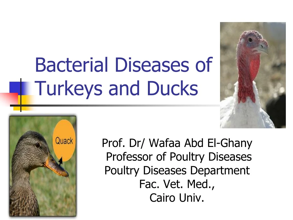
Bacterial Diseases of Turkeys and Ducks Prof. Dr/ Wafaa Abd El-Ghany Professor of Poultry Diseases Poultry Diseases Department Fac. Vet. Med., Cairo Univ.
Bacterial diseases of Turkeys • Mycoplasma gallisepticum (MG). • Mycoplasma meleagridis (MM). • Maycoplasma synoviae (MS). • Mycoplasma iowae (MI). • Turkey coryza (Bordetellosis). • Ornithobacterium rhinotracheal (ORT). • Erysiplas infection. • Avian chlamydiosis. • Fowl cholera. • Avian salmonellosis (Paratyphoid infection, Arizona disease and Pullorum disease). • E.coli infection. • Clostridial infections. • Staphylococcus and Streptococcus infections. • Avian Tuberculosis
Bacterial diseases of Ducks • Rimerella anatipestifer • Botulism (Western duck sickness, Limber neck) • Paratyphoid infection. • Avian chlamydiosis • Mycoplasma immitans & ansaris • Avian spiroketosis • E. coli infection • Staphylococcus and Streptococcus infections
AVIAN CHLAMYDIOSIS (ORNITHOSIS)
Avian chlamydiosis • Avian chlamydiosis is acute fatal or chronic infectious zonootic respiratory disease of domestic, wild and migratory birds. • The disease is characterized by nasal and ocular discharge, diarrhea, loss of weight, drop in egg production and the mortality rate reached to 5-30%. • Presence of intracytoplasmic elementary bodies (LCL) is characteristic to the organism.
Giemsa stain: intracellular chlamydial inclusion (reddish-purple).
Infection & Transmission of Chlamydiosis • Chlamydia Psittaci infection occur horizontally either by inhalation of infected dust or droplet or by ingestion of contaminated feed and water with infected droppings. • There is an evidence of low percentage of vertical transmission through eggs (transoverian) in ducks and sea gulls. • Chronic carriers transmit the organism without signs. • Biting of insects as ticks, lice and mites. • Wild birds are important in transmission.
Signs of chlamydiosis In turkeys: I Acute epidemic Form (toxigenic highly virulent strain): Clinical signs appear as systemic fatal infection with: • Sudden death of 5-30%. • Fever and anorexia. • Nasal and ocular discharge, swollen eye lids, conjunctivitis and sinusitis. • Off food and loss of weight (emaciation). • Yellow green gelatinous diarrhea. • Rapid drop in egg production (40-50% decline) • Morbidity rate may be reach 50-80%.
Signs of chlamydiosis In ducks and geese (water fowl): • In duckling there are tremors, trembling and staggered gait (incoordination) • Ocular and nasal discharge. • Greenish watery Diarrhea • Inappetance • Emaciation, convulsion and death. • Morbidity rate 10-80%. • Mortality rate 1-30% (depending on the age, health status and secondary infection with salmonella species).
Lesions of chlamydiosis • Congestion of the visceral organs. • Congestion and pneumonia of the lungs. • All the body cavities are filed with fibrinous exudates. • Fibrinous pericarditis, perihepatitis, and airsacculitis and peritonitis. • Catarrhal enteritis. • Liver and spleen are enlarged, dark and covered with gray white foci.
Fibrinous pericarditis,liver hepatomegaly and congestion. Liver necrosis fibrinous pericarditis. Fibrinous pericarditis
Caseous pericarditis. Lungs congested and fibrinous exudate in the pleural cavity Necrosis and congestion of spleen
Prehepatitis Enlarged congested liver Airsacculitis Chronic or Lesions of Prolonged CP infection
A. Thickened abdominal airsacs totally covered with fibrin plaques . B. Serous fluid and fibrin in the pericardial sac. C. Severe hepatomegaly.
Diagnosis of chlamydiosis • Detection of dark purple, blue or pink (acc. To the stain) I/Cytoplasmic inclusion (LCL) bodies in stained smear (from nasal or ocular exudates, serous membranes, trachea, lungs, pancreas, liver, spleen, heart, faecal and cloacal swabs) in different stages of elementary body. • Tissue culture. • Embryonated chicken eggs. • Laboratory animal inoculation: Intra peritoneal or intra cranial or intra nasal inoculation of 3-4 weeks old mice (3-6 mice) could resulted in death of mice after 5-7 days post inoculation. Intra peritoneal inoculation resulted in peritonitis with accumulation of fibrinous exudate in the peritoneal cavity with splenomegaly.
Prevention of chlamydiosis • Different avian species and ages must be reared away from each other. • Newly purchased (imported) birds, especially pets must be quarantined for 35 days (if positive discarded or treated with tetracyclines till recovery). • Prevent the introduction of birds from the enzootic areas. • Avoid the contact with the free living wild birds. • Thorough cleaning and disinfection (iodophors or formaldhydes). • Regular testing of the birds using serological tests. • Restrict the movement of people and visitors. • Trials for vaccine preparation.
Control of chlamydiosis • Oxytetracyclin (15mg/kg bwt) in the drinking water for a period for 5 days. • Chlorotetracyclin. (treatment may be extended to 15-45 days in severe infection). • Usage of combined antibiotics to control other associated infections as Salmonellae and E.coli (quinolones like cipro, enro, and danofloxacin) for 3-5 days in the drinking water.
Avian spiroketosis • Acute septicaemic and chronic disease. • The bacterium Borrelia anserina infects chickens, turkey, geese, ducks, pheasants, grouse and canaries with morbidity and mortality up to 100%. • It is transmitted by arthropods, e.g. Argas persicus, and occasionally by infected faeces. The bacterium is poorly resistant outside host but may be carried by Argas persicus for 430 days.
Signs & lesions of avian spiroketosis • Depression and thirst • Cyanosis. • Often diarrhoea with excessive urates. • Weakness and progressive paralysis. • Drops in egg production may be seen in both systemic and intestinal forms Post-mortem lesions • Marked splenomegaly. • Spleen mottled with ecchymotic haemorrhages. • Liver enlarged with small haemorrhages. • Necrotic foci. • Mucoid enteritis.
Prevention & Control of avian spiroketosis Treatment • Various antibiotics including penicillin. Prevention • Control vectors. • Vaccines in some countries.
Cause Age group affected Transmission Forms of the disease Escherichia coli All types of avian and all ages Fecal oral route, transovarial, contamination of the egg shell via fecal material from hen Colisepticaemia Airsac disease Chronic forms (arthritis, omphalitis, panophthalmitis, salpingitis, enteritis, coligranuloma and SHS). E. coli
Severely congested liver Foci of necrosis on liver
GANGARINOUS DERMATITIS (GD) GAS OEDEMA DISEASE WING ROT
Gangrenous dermatitis • Gangrenous (necrotic) dermatitis in broilers and turkeys between 3-7 weeks old. • It is caused by a bacteria- Clostridium septicum, Escherichia coli, Staphylococcus aureus. • Disease often occurs in birds which are already immunosuppressed due to a prior infection (Chicken anaemia, Infectious Bursal Disease) or mycotoxin ingestion. • Transmission is by contact with infected wet, caked litter. The disease often occurs in immunosuppressed birds.
Signs of GD • Loss of feathers • Pale combs and wattles, depression • Incoordination • Leg weakness and ataxia (can’t move) can be seen • Mortality is low, but dead birds decompose quickly.
Lesions of GD • Congestion, haemorrhage and necrosis of skin with intro lesion bacteria under the microscope. • In turkeys with cellulitis of the tail (bubbly tail), edema and vesicle-like lesions were present laterally and ventrally around the tail. • Tail feathers were soft, blood-filled, and broken.
Necrosis of the wattle Dark and moist skin of the breast Affected leg and toes. Necrosis of the abdomen skin.
Prevention & Control of GD Prevention • Clean out house and add new litter to prevent the disease. • Medicate in starter feed Flavomycin, Virginamyciin, Bacitrcin and CTC can reduce bacteria. • Proper vaccination against IBDV, CAV and MDV prevent mycotoxin formation in the feed, and eliminate ALV in the breeders to prevent immunosuppression. Treatment • Erythromycin, penicillin in the feed to treat signs. Chlortetracycine, oxytetracycline, copper sulphate can be added to the water to reduce morbidity.
ULCERATIVE ENERITIS (UE) Quail disease
Ulcerative Enteritis • The agent involved in the aetiology of this disease is clostridium colinum, which is spore-forming, gram-positive, aerobic and non-motile. Mode of transmission • Vectors are faeces, soil and litter containing the bacteria. • It often accompanies coccidiosis in broilers.
Signs & lesions of UE • High mortality, watery diarrhoea, ruffled feathers, dull, listlessness, increased thirst, emaciation and atrophy of pectoral muscles can occur. Postmortem lesions • Yellow irregular ulcers on small intestine and caeca, hemorrhagic enteritis are seen. • Enlarged haemorrhagic necrotic spleen, light yellow mottling of liver and crop filled with water may occur.
Prevention & Control of UE Prevention • Improved sanitation. Adding salt to the soil (500 lbs of salt/house) may kill spores. Raising birds on wire and/or feeding bacitracin at 50-100 g/t will prevent the disease. Treatment • NF-180 (50-100 g/t), streptomycin (60 g/t) and chlortetracycline, vitamins and minerals in water and/or lincomycin 2 g/t will reduce the signs. • Remove dead birds and feed bacitracin (200 g/t)