Download

1 / 94
1k likes | 1.4k Views
Bacterial Diseases. Victor Politi,M.D., FACP, Medical Director, SVCMC School of Allied Health Professions, Physician Assistant Program. Introduction. Bacteria consist of only a single cell Bacteria fall into a category of life called the Prokaryotes

E N D
Bacterial Diseases Victor Politi,M.D., FACP, Medical Director, SVCMC School of Allied Health Professions, Physician Assistant Program
Introduction • Bacteria consist of only a single cell • Bacteria fall into a category of life called the Prokaryotes • There are thousands of species of bacteria, but all of them are basically one of three different shapes.
Classification of Bacteria • Until recently classification has done on the basis of such traits as: • shape • bacilli: rod-shaped • cocci: spherical • spirilla: curved walls • ability to form spores • method of energy production (glycolysis for anerobes, cellular respiration for aerobes • nutritional requirements • reaction to the Gram stain.
Classification of Bacteria • The Gram stain is named after the 19th century Danish bacteriologist who developed it. • The bacterial cells are first stained with a purple dye called crystal violet. • Then the preparation is treated with alcohol or acetone. • This washes the stain out of gram-negative cells. • To see them now requires the use of a counterstain of a different color (e.g., the pink of safranin). • Bacteria that are not decolorized by the alcohol/acetone wash are gram-positive
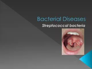
Gram Positive Bacteria • I-Gram Positive Cocci • A-Streptococcus (e.g. streptococcus Pneumoniae) • B-Staphylococcus (e.g. Staph. aureus) • C-Enterococcus (Previously Group D Strep.) • II-Gram Positive Rods • A-Corynebacteria: Corynebacterium diphtheria • B-Listeria monocytogenes • C-Bacillus anthracis (Anthrax) • D-Erysipelothrix rhusiopathiae • III-Gram Positive Branching Organisms • A-Actinomycetes
Gram Positive Cocci • I-Beta-hemolytic Streptococcus (Lancefield Groups) • - Group A Streptococcus (Streptococcus Pyogenes) • - Group B Streptococcua (Streptococcus agalactiae) • - Group C Streptococcus • - Group G Streptococcus • II-Alpha-hemolytic Streptococcus • - Streptococcus Pneumoniae (Pneumococcus) - Viridans streptococcus (bacterial endocarditis) • III-Non-hemolytic Streptococcus • - Streptococcus faecalis (Group D) • - Certain members of Groups B, C, D, H, and O
Strep throat is caused by group A Streptococcus bacteria. These bacteria are spread through direct contact with mucus from the nose or throat of persons who are infected, or through contact with infected wounds or sores on the skin
Group B Streptococcus (Streptococcus agalactiae) • Epidemiology • Most common US cause of neonatal sepsis and meningitis • Incidence • Overall: 2 to 4 per 1000 live births • Invasive: 1.8 per 1000 live births • Primarily occurs in newborns • Very rare after 5 months of age
Group B Streptococcus(Streptococcus agalactiae) • Pathophysiology • Group B Beta-hemolytic streptococcus infection • Perinatal transmission • Delivery via a birth canal colonized with GBS • Incidence of U.S. vaginal GBS colonization: 15-20% • Onset of infection (Mean onset 20 hours of life) • Early onset neonatal disease (<6 days of life in 80%) • Sepsis • Pneumonia • Late onset neonatal disease of sepsis or mengitis
Group B Streptococcus (Streptococcus agalactiae) • Labs: Maternal Screening • GBS Culture • Management • Sepsis (treat for 10-14 days) • Pencillin G 200,000 units/kg/day divided q4-6 hours • Meningitis (treat for 14-21 days) • Penicillin G 400,000 units/kg/day divided q2-4 hours • Prevention • Perinatal Group B Streptococcus Prophylaxis • Prognosis • Mortality 10-40%
Streptococcus Pneumoniae (Pneumococcus) • Epidemiology • Most common cause of community acquired pneumonia • Classic Symptoms • Shaking rigors • Fever • Purulent sputum • Rust colored • Pleuritic chest pain • Dyspnea • Chest splinting
Alpha-hemolytic Streptococcus • Lab • CBC • WBC elevated with left shift • Gram stain • Gram positive encapsulated organisms • Elongated lancet shaped diplococci • Blood Culture • Positive in only 33% of cases • Sputum culture • Positive in only 40% of pneumococcal pneumonias • Radiology • Chest X-ray • Lobar consolidation (often lower lobe) • patchy infiltrates
Management • Increasing Pencillin Resistance • Penicillin Sensitive • Ampicilin IV or Amoxicillin PO • Erythomycin • Azithromycin • Clarithromycin • Penicillin G IV • Doxycycline • Oral second generation cephalosporin • Parenteral third generation cephalosporin
Management • High-Level Penicillin Resistance • Broad spectrum Fluoroquinolone • Levofloxacin • Gatifloxacin • Grepafloxacin • Moxifloxacin • Sparfloxacin • Parenteral third generation Cephalosporin • High dose Ampicillin • Vancomycin IV with or without Rifampin
Gram Positive Cocci • Organisms • -Staphylococcus aureus • -Staphylococcus epidermidis
Enterococcus • I-Characteristics • Gram Positive Cocci • Previously defined as Group D Streptococcus II-Organisms • Enterococcus faecalis • Enterococcus faecium
Corynebacterium • Epidemiology • Rare in United States due to Immunization (DTP, DTaP) • However 20% of adults may be inadequate immune status • Ongoing epidemic in the former USSR • Etiology • Corynebacterium Diphtheriae
Corynebacterium • Symptoms • sore throat • dysphagia • Weakness • Malaise
Corynebacterium • Signs • Toxic appearance • fever • Tachycardia (out of proportion to fever) • Pharyngeal erythema • Gray-white tenacious exudate or "membrane" • Occurs at tonsillar pillars and posterior pharynx • Leaves focal hemorrhagic raw surface when removed • Cervical lymphadenopathy
Differential Dx • Vincent's Angina (trench mouth) • Also shows pseudomembrane formation • Pharyngitis • Labs • CBC • Leukocytosis • Throat culture (+ for corynebacterium org.) • Management • Diphtheria antitoxin • Erythromycin • 20-25 mg/kg q12 hours IV for 7-14 days • Prevention • DTP/DTaP vaccination
Bacillus anthracis (Anthrax) • Etiology • Bacillus anthracis • Transmission • Contact with hides of infected animals • Cattle • Sheep • Camels • Antelopes • Ingestion of contaminated meat • Inhalation of spores • Infective aerosol dose: 8,000-50,000 spores • Spores may remain viable in soil for >40 years • No transmission person to person
Bacillus anthracis (Anthrax) • Symptoms and Signs: Cutaneous ("Malignant Pustule") • Inoculation at site of broken skin • Painless pruritic pustules develop at inoculation site • Begins as erythematous papule on exposed skin • Vesiculates and then ulcerates within 1-2 days • Surrounded by a ring of non-tender Brawny edema • Black eschar may form
Bacillus anthracis (Anthrax) • Symptoms and Signs: Inhalation Anthrax • Malaise • Regional lymphadenopathy • Two phases • Initial Phase • Viral upper respiratory symptoms • rhinorrhea • pharyngitis • Later Phase • dyspnea and hemoptysis during dissemination
Acute GI type symptoms Hematemesis Severe diarrhea Differential Diagnosis Cutaneous Anthrax Spider bite Ecthyma gangrenosum Ulceroglandular tularemia Plague Staph. Or strep. cellulitis Inhalational Anthrax Community acquired pneumonia (late phase anthrax) Mycoplasma pneumonia (early phase anthrax) Influenza (early phase anthrax) Legionnaires' Disease Psittacosis tularemia Q fever Viral pneumonia Histoplasmosis Coccidiodomycosis Symptoms and Signs:
Bacillus anthracis (Anthrax) • Labs • Rapid ELISA test now available • Cultures • Blood culture (high sensitivity) • Cultures of Vomitus or feces (Intestinal Anthrax) • CSF culture (Inhalational Anthrax) • Nasal Swab (Epidemiologic tool to identify outbreak) • Sputum culture (Inhalational Anthrax) • Vesicular fluid (Cutaneous Anthrax) • Gram stain - blood or vesicular fluid from lesion • Gram positive bacilli • CBC • Neutrophilic leukocytosis in severe cases • Radiology: • Chest x-ray - Widened Mediastinum (hemorrhagic mediastinitis
Antibiotic course: 60 days Empiric Treatment Cipro Adults: 400 mg IV q12 hours Children: 20-30 mg/kg/day IV divided q12 hours Levofloxacin Adults: 500 mg IV q24 hours Specific Treatment for confirmed anthrax Adults Pencillin G 4 MU IV q4 hours or Doxycycline 200 mg IV, then 100 mg IV q12 hours Children > age 12 same as adults Children < age 12 Penicillin G 50,000 U/kg IV q6 hours Management: Antibiotics
Postexposure prophylaxis • Concurrently begin vaccination • Continue antibiotics for 60 days • Ciprofloxacin • Adults: 500 mg PO bid • Children: 20-30 mg/kg/day divided bid up to 1g/day • Amoxicillin • Adults: 500 mg PO tid • Children: 40 mg/kg up to 500 mg PO tid • Doxycycline • Adults: 100 mg PO bid • Children over age 8: 5 mg/kg/day divided q12 hours
Anthrax • Course • Incubation: 4-6 days • Duration of illness: 3-5 days • Prognosis • Inhalation Anthrax (inhaled spores) • Untreated: 95% mortality • Treated: 80% mortality • Cutaneous Anthrax (skin contact) • Untreated: 20% mortality • Treated: Rare mortality • Intestinal Anthrax (ingested contaminated meat) • Mortality 25 to 60%
Prevention • Anthrax Vaccine 93% effective • Initial: 0, 2, and 4 weeks • Next: 6, 12, 18 months and then annually • Postexposure Prophylaxis as above • Empiric prophylaxis for any suspected exposure • Best prognosis with antibiotics prior to symptoms
Gram Negative • Gram Negative Rods • Anaerobes • Bacteroidaceae (e.g. Bacteroides fragilis) • Facultative Anaerobes (enteric/nonenteric) • Enterobacteriaceae (e.g. Escherichia coli) • Vibrionaceae (e.g. Vibrio Cholerae) • Pasturella,Brucella,Yersinia • Aerobes • Pseudomonadaceae (e.g. Pseudomonas aeruginosa)
Facultative Anaerobes • Enterobacteriaceae (e.g. E. coli) • Vibrionaceae (e.g. Vibrio Cholerae) • Salmonella,Shigella,Klebsiella,Proteus • GI pathogens !!!!! • non-enteric Pasturella,Brucella,Yersinia • Francisella,Hemophilus,Bordetella
Enterobacteriaceae • Characteristics • Facultative Anaerobic Gram negative rods • EKP Gram negative bacteria • Escherichia coli • Klebsiella • Proteus
Vibrionaceae • Characteristics • Facultative Anaerobic gram negative rods • Vibrio Cholerae • Vibrio parahaemolyticus • Genus: Aeromonas (motile with single polar flagellum)
Vibrionaceae • Genus: Campylobacter (motile with single polar flagellum) • Campylobacter jejuni • Genus: Helicobacter (motile with multiple flagella) • Helicobacter Pylori
Pasteurellaceae • Characteristics • Facultative Anaerobic gram negative rods • Genus: Pasteurella • Pasteurella multocida
Pasteurellaceae • Genus: Haemophilus (coccobacilli) • Haemophilus Influenzae • Haemophilus aegyptius • Haemophilus ducrei
Gram Negative Rod • Aerobes • Pseudomonadaceae (e.g. Pseudomonas aeruginosa) • Brucella • Legionellaceae
Pseudomonadaceae • Characteristics • Aerobic Gram Negative Rod • Family: Pseudomonadaceae • Pseudomonas aeruginosa • Pseudomonas mallei (Glanders)
Gram Negative Rod Aerobic • Family: Legionellaceae • Legionella pneumophila
Pathophysiology Aerobic, intracellular, Gram negative rod Virulent organism More severe disease than other atypical pneumonia Transmission Optimal conditions for growth Temperature: 89 to 113 F water Stagnant water Transmission Waterborne Freshwater or moist soil near ponds Air conditioning Condensers Cooling towers Respiratory therapy equipment Showers or water faucets Whirlpools Incubation Two to ten days Legionellaceae
Symptoms Prodrome for 12-48 hours Malaise Myalgia HA Symptoms for 2-3 days Fever to 40.5 C persists for 8-10 days GI symptoms- 20-40% of cases Nausea/vomiting Diarrhea Later Symptoms: Cough Minimal to no sputum production Slightly blood tinged sputum Signs Severe respiratory distress Confusion Disorientation Legionellaceae
Legionella pneumophila • Complications • Respiratory failure (20-40% of cases) • Extrapulmonary complications • Myocarditis/pericarditis • Prosthetic valve endocarditis • Glmoerulonephritis • Pancreatitis • Peritonitis
Legionella pneumophila • Radiology: chest x-ray • Small pleural effusions • Unilateral parenchymal infiltrates • Round, fluffy opacities • Spread contiguously to other lobes • Progresses to dense consolidation • Progresses to bilateral infiltrates