DNA EXTRACTION
450 likes | 701 Views
DNA EXTRACTION. Have you done DNA extractions with your class?. 1865 presented paper Experiments on Plant Hybridization,. Johann Friedrich Miescher 1844 -1895. 1869: Characterizes a new substance in pus (June 15, 1866-August 23, 1866).

DNA EXTRACTION
E N D
Presentation Transcript
Johann Friedrich Miescher 1844 -1895 1869: Characterizes a new substance in pus (June 15, 1866-August 23, 1866). “… the substance was derived from the nucleus of the cell. Hence, we call it nuclein.”
Pál Plósz (1871) verified the presence of nuclein in the nucleated erythrocytes of birds and reptiles and its absence from the erythrocytes of mammals, which are devoid of a nucleus.
1881: (Ludwig Karl Martin Leonhard)Albrecht Kossel determines that nucleic acid is composed of four bases. Untersuchungen über die Nukleine und ihre Spaltungsprodukte (Investigations into the nucleins and their cleavage products) 1910
1893: Albrecht Kossel determines that nucleic acid is composed of four bases. 2 purines: 2 pyrimidines: adenine (A) guanine (G) cytosine (C) thymine (T)
1889: Richard Altmann finds that nuclein is acidic and renames it nucleic acid (nucleïnsäure). Ueber Nucleinsäuren. Archiv für Anatomie und Physiologie. Physiologische Abteilung. Leipzig, 1889, 524-536.
Phoebus Aaron (Theodore) Levene (1869-1940) 1909: Phoebus Levene discovers that DNA is made of 3 basic components: a sugar, an acid, and an organic base.
1944: Oswald Avery, Colin MacLeod, and Maclyn McCarty establish that Griffith's transforming principle is DNA, and suggest that it may function as the genetic material. Avery, O. T., MacLeod, C. M. & McCarty, M. 1944. Studies of the chemical nature of the substance inducing transformation of pneumococcal types. Induction of transformation by a desoxyribonucleic acid fraction isolated from Pneumococcus Type III. J. Exp. Med.79:137-158.
1949: Roger Vendrely, Colette Vendrely, and André Boivin find half as much DNA in the nuclei of sex cells as they find in body cells, thus paralleling the reduction in the number of chromosomes, making DNA look like the genetic material.
1951 X-ray diffraction of DNA Rosalind Franklin
1952: Alfred D. Hershey & Martha Chase DNA is the genetic material
Martha Chase Awarded a blender (?) Alfred D. Hershey Awarded the Nobel Prize
“We have formulated a structure for the nucleic acids… The structure involves three intertwined helical polynucleotide chains. Each chain… has approximately twenty-four nucleotide residues in seven turns of the helix. The helixes form a right-handed screw. The phosphate groups are closely packed about the axis… with the pentose residues… and the purine and pyrimidine groups projecting radially...” Linus Pauling & Robert B. Corey (Nature, 1953, 171:346).
Pauling, L. and Corey, R. B. 1953. A proposed structure for the nucleic acids Proc. Natl. Acad. Sci. USA 39:84-97.
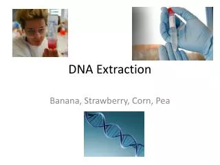
What are the essential components of a DNA extraction Procedure? Maximize DNA recovery Remove inhibitors Remove or inhibit nucleases Maximize the quality of DNA
How Much DNA Do We Need? • The PCR reactions call for on average 1 ng of DNA (single or double stranded). • Many of the commercially available kits are sensitive below 1 ng of DNA (100-250 pg).
Basic steps for DNA extraction • Breaking the cells open, commonly referred to as cell disruption or cell lysis, to expose the DNA within. This is commonly achieved by grinding, sonicating or treating the sample with lysis buffer . 2. Removing membrane lipids by adding a detergent.
Lysis Buffer: 50 ml Lysis Buffer 5 mM EDTA pH 8.0 0.5 M EDTA = 500 ul 200 mM NaCL 5M NaCl = 4 mL 100 mM Tris pH 8.0 1 M Tris-HCL ph 8= 5 mL 0.2% SDS Sodium dodecyl Sulfate 10% SDS = 1 mL Water MQ water = 39.5 mL Add prior to 55C o/n icubation 0.4 mg/ml Proteinase K 20 mg/mL Prot K = 6 ul per 300ul of Lysis Buffer
Detergents Chaotropic salts CTAB Purposes of the Extraction Buffer 1. Dissolve cellular membranes 2. Inactivation of DNase and Rnase 3. Assist in the removal of contaminants Detergents Metal chelators Reducing agents Salts CTAB PVP
Extraction/Precipitation Method Use of Detergents to Lyse Cells: Mixed micelle Detergent molecules Plasma membrane (phospholipid bilayer) + SDS
3. Removing proteins by adding a protease (optional but almost always done). 4. Precipitating the DNA with an alcohol — usually ice-cold ethanol or isopropanol. Since DNA is insoluble in these alcohols, it will aggregate together, giving a pellet upon centrifugation. This step also removes alcohol-soluble salt.
Most Commonly used DNA Extraction Procedures • Organic (Phenol-Chloroform) Extraction • Non-Organic (Proteinase K and Salting out) • Qiagen (anion exchange resin) The method utilized may be sample dependant, technique dependant, or analyst preference
EXTRACTION • Perhaps the most basic of all procedures in genetic engineering is the purification of DNA. The key step, the removal of proteins, can often be carried out simply by extracting aqueous solutions of nucleic acids with phenol and/or chloroform.
ORGANIC EXTRACTION REAGENTS • Cell Lysis Buffer - Non-ionic detergent , Salt, Buffer, EDTA designed to lyse outer cell membrane, but will not break down nuclear membrane. • EDTA (Ethylenediaminetetraacetic disodium salt) is a chelating agent of divalent cations such as Mg2+. Mg2+is a cofactor for Dnase nucleases. If the Mg2+is bound up by EDTA, nucleases are inactivated.
ORGANIC EXTRACTION REAGENTS • Proteinase K - it is usual to remove most of the protein by digesting with proteolytic enzymes such as proteinase K, which are active against a broad spectrum of native proteins, before extracting with organic solvents. Protienase K is approximately 10 fold more active on denatured protein. Proteins can be denatured by SDS or by heat.
ORGANIC EXTRACTION REAGENTS • Phenol/Chlorform - The standard way to remove proteins from nucleic acids solutions is to extract once with phenol, once with a 1:1 mixture of phenol and chloroform, and once with chloroform. This procedure takes advantage of the fact that deproteinization is more efficient when two different organic solvents are used instead of one. • Also, the final extraction with chloroform removes any lingering traces of phenol from the nucleic acid preparation. • Phenol is highly corrosive and can cause severe burns.
Extraction/Precipitation Method Step 1:Disruption of cell walls by grinding Step 1+2:mechanical disruption and homogenization in extraction buffer Grind sample into a fine powder to shear cell walls and membranes Step 2:Lysis of cells in extraction buffer A homogenizer allows cells to be mechanically disrupted within the extraction buffer Mix thoroughly with extraction buffer to dissolve cell membranes and inhibit nuclease activity Crude lysate
Extraction/Precipitation Method Step 3:Organic extraction Mix thoroughly with an equal volume of organic solvent Aqueous Centrifuge Collect aqueous phase e.g. phenol, chloroform, or phenol:chloroform Interphase Organic Perform additional extractions for increased purity Crude lysate containing nucleic acids and other cell constituents The aqueous phase contains water-soluble molecules, including nucleic acids. Proteins and lipids become trapped in the organic phase, and are thus separated away. Insoluble plant debris become trapped in the interphase between the two layers
Extraction/Precipitation Method Step 4:Nucleic Acid Precipitation Before After After Supernatant 70% EtOH Centrifuge Wash Centrifuge Pellet Dissolve pellet (H2O, TE, etc.) • Pellet down nucleic acids. • Pellet down nucleic acids. • Wash pellet with 70% ethanol to remove residual salts and other contaminants. • Pellet down nucleic acids. • Wash pellet with 70% ethanol to remove residual salts and other contaminants. • Discard ethanol and allow pellet to dry. Add alcohol and salt to precipitate nucleic acids from the aqueous fraction
Concentrating DNA byAlcohol Precipitation • The most widely used method for concentrating DNA is precipitation with ethanol. The precipitate of nucleic acid, forms in the presence of moderate concentrations of monovalent cations (Salt, such as Na+), is recovered by centrifugation and redissolved in an appropriate buffer such as TE. • The technique is rapid and is quantitative even with nanogram amounts of DNA.
Concentrating DNAAlcohol Precipitation • The four critical variables are the purity of the DNA, its molecular weight, its concentration, and the speed at which it is pelleted. • DNA a concentrations as low as 20 ng/ml will form a precipitate that can be quantitatively recovered. • Typically 2 volumes of ice cold ethanol are added to precipitate the DNA.
Concentrating DNAAlcohol Precipitation • Very short DNA molecules (<200 bp) are precipitated inefficiently by ethanol. • The optimum pelleting conditions depend on the DNA concentration. Relatively vigorous microcentrifuge steps such as 15 minutes at or below room temperature at 12,000 rpm are designed to minimized the loss of DNA from samples with yields in the range of a few micrograms or less.
Concentrating DNAAlcohol Precipitation • Solutes that may be trapped in the precipitate may be removed by washing the DNA pellet with a solution of 70% ethanol. To make certain that no DNA is lost during washing, add 70% ethanol until the tube is 2/3 full. Vortex briefly, and recentrifuge. After the 70% ethanol wash, the pellet does not adhere tightly to the wall of thetube, so great care must be taken when removing the supernatant.
Concentrating DNAAlcohol Precipitation • Isopropanol (1 volume) may be used in place of ethanol (2 volumes) to precipitate DNA. Precipitation with isopropanol has the advantage that the volume of liquid to be centrifuged is smaller. • Isopropanol is less volatile than ethanol and it is more difficult to remove the last traces; moreover, solutes such sodium chloride are more easily coprecipitated with DNA when isopropanol is used.
Resuspension and Storage of DNA • TE Buffer - Tris-EDTA Buffer: 10 mM Tris-HCl pH 8.0, 1 mM EDTA, or TE-4 which is 10 mM Tris, 0.1 mM EDTA. DNA is resuspended and stored in TE buffer. DNA must be stored in a slightly basis buffer to prevent depurination, and the EDTA chelates any Mg2+ helping to inactivate DNases.
DNA can be stored at 4oC for extended periods, however for long term storage, - 20oC is preferable. • Avoid repetitive freeze thawing of DNA, since this can cause degradation.
Checking for DNA genomic DNA Running nucleic acid sample through an agarose gel is a common method for examining the extent of DNA degradation. Good quality DNA should migrate as a high molecular weight band, with little or no evidence of smearing. RNA (degraded)
Nucleic Acid Analysis via UV Spectrophotometry DNA Absorption Spectra By measuring the amount of light absorbed by your sample at specific wavelengths, it is possible to estimate the concentration of DNA and RNA. Nucleic acids have an absorption peak of 1 OD at ~260nm. [dsDNA] ≈ A260 x (50 µg/mL) [ssDNA] ≈ A260 x (33 µg/mL) [ssRNA] ≈ A260 x (40 µg/mL)
How pure is nucleic acid sample? Nucleic acids strongly absorb at 260 nm and less strongly at 280 nm while proteins do the opposite. The A260/A280 ratio is ~1.8 for dsDNA, and ~2.0 for ssRNA. Ratios lower than 1.7 usually indicate significant protein contamination. The A260/A230 ratio of DNA and RNA should be roughly equal to its A260/A280 ratio (and therefore ≥ 1.8). Lower ratios may indicate contamination by organic compounds (e.g. phenol, alcohol, or carbohydrates).