Download
1 / 32
390 likes | 896 Views
Medical Exposure. Dr. Nabil Maalej. Exposure To Radiation. Radiation Dose. Specification of the radiation absorbed dose in Gy or rad is inadequate for complete and accurate assessment of radiation hazard. Dose Modifying Factors. The type of radiation involved (w R )

E N D
Medical Exposure Dr. Nabil Maalej
Radiation Dose Specification of the radiation absorbed dose in Gy or rad is inadequate for complete and accurate assessment of radiation hazard
Dose Modifying Factors • The type of radiation involved (wR) • The part of the body exposed (wT) :the active blood forming organs, the gonads, and the lens of the eye are specifically sensitive • The time span over which the radiation dose is delivered • The age of the exposed individual: the developing fetus is especially sensitive
Dose Equivalent • The dose equivalent is the absorbed dose in tissue multiplied by a quality factor wR : • HT,R = wR DT,R • The SI units for dose equivalent is the Sievert = 1 J/kg for wR =1 • 1 Sievert = 100 rem
Effective Dose Effective Dose is the summation of tissue equivalent doses each multiplied by the tissue weighing factor:
X-Rays • Diagnostics radiology is necessary to visualize human anatomy and detect disease • Good image quality and low patient doses are frequently conflicting requirements • The medical practitioner should keep the radiation exposure As Low As Reasonably Achievable (ALARA) to realize the benefit of imaging while minimizing dose to the patient.
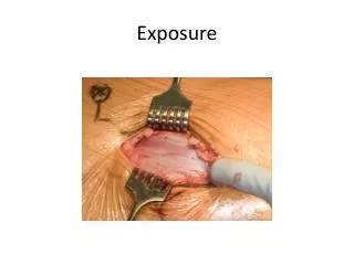
Exposure • Exposure refers to the amount of ionization of air caused by x-ray and -ray beams • Exposure E = Q/m • The unit is the Roentgen (R): the ionization liberating an amount of charge equal to 2.58 X 10⁻⁴ coulombs/kg of air • The SI units is C/kg = 3876 R • Exposure can be measured using ionization chambers
Exposure Variation With X-ray Machine Settings • Exposure (kVp)2 (1/d)2 (mAs) • kVp is the applied voltage, d is the distance from the focal spot, and mAs is the applied current multiplied by the time • Typically, for d = 1 m and kVp = 100 and a 2mm Aluminum filtration of the beam, the Exposure is 10 mR/mAs
Example 1 Estimate the exposure at a distance d = 2 m for 5 mAs and 150 kVp, assume 2 mm Al filtration?
X ray Dose to a Patient • If the exposure is measured at a location , the absorbed dose in rads that would be delivered to a person at that location can be estimated by means of the f factor • For practical purposes the exposure level in roentgens is approximately equal to to the absorbed dose in rads which is equal to the dose equivalent in rems
X ray Dose to a Patient • Surface Doses can also be conveniently measured using TLDs • Doses to inaccessible organs can be measured using body equivalent phantoms • Mathematical phantoms such as MIRD-5 and mathematical calculations such as Monte Carlo can be used to estimate doses
Reduction of Exposure • The medical practitioner should keep the radiation exposure As Low As Reasonably Achievable (ALARA) to realize the benefit of imaging while minimizing dose to the patient. • Use of contrast media such as barium sulfate and organic iodine • Use digital radiography to capture and store images in digital form • Digital Subtraction Angiography
Nuclear Medicine • There is a rapid expansion in the diagnostic use of radio-pharmaceuticals • Some common procedures are bone, thyroid, hart, lung, liver and kidney scans • 99mTc is the radionuclide of choice in 75% of nuclear medicine procedures
Maximum Permissible Dose • The maximum permissible dose (MPD) to the whole body for radiation workers should not exceed 50 mSv per year as recommended by ICRP (International Commission of Radiological Protection) and NCRP • The Nuclear Regulatory Commission NRC recommends that and individual lifetime exposure should not exceed (N-18) X 50 mSv, where N is the age in years
Radiation Safety • Beside observing the MPD the rule is to keep radiation as low as reasonably achievable (ALARA) • ALARA can be applied to the handling of radiation sources, to storage and shielding techniques, and to the design and layout of the laboratory
Exposure Rate Constant • The exposure rate constants (R.cm2/mCi.hr) for a radionuclide is the exposure due to -rays and x-rays in R.hr, at a distance of 1 cm from an unshielded 1 mCi source of that radionuclide • Radiation exposure levels caused by -ray and x-ray emitters can be estimated from exposure rate constants
External Dose Calculation • The exposure rate E(R/hr) at a distance d(cm) from a source of activity A(mCi) and an exposure rate constants (R.cm2/mCi.hr) E = A /d2 • Because -rays and x-rays are emitted isotropically exposure levels decrease as the square of distance from the source
Example Calculate the exposure rate at 10 cm and 300 cm distances from a syringe containing 30 mCi of 99mTc? The exposure rate constant for 99mTc is 0.6 (R.cm2/mCi.hr).
Answer At d=10 cm, E = A /d2= 30(mCi) X 0.6 (R.cm2/mCi.hr)/(10 cm2) = 0.18 R/hr = 180 mR/hr And at d=10 cm, E = 30(mCi) X 0.6 (R.cm2/mCi.hr)/(300 cm2) = 0.0002 R/hr = 0.2 mR/hr This illustrates the strong effect of distance on radiation exposure
Reduction of Exposure from External Sources The basic principle of reducing doses from external exposure are described by the Time, Distance, Shielding (TDS) rules: • Decrease the time of exposure • Increase the distance from the source • Use shielding where necessary
Effective Shielding Use lead boxes for storage, lead lined syringes, lead aprons, lead bricks for lining storage area, leaded glass for viewing
Prevention of Internal Exposure • No eating, drinking or smoking in “hot labs” • Wear lab coats and gloves • No food storage in hot areas • Absolutely no pipetting • Wash hands • Perform work on absorbent pads and in ventilated hoods • Promptly and appropriately dispose of radioactive waste • Appropriately store radioactive materials • Use personnel dosimeters and laboratory monitors
Conclusion • The exposure from natural radiation is on the average higher than the exposure from medical radiological procedures • In order to minimize damage from radiation it is necessary to observe the maximum allowable limits and to keep exposure As Low As Reasonably Achievable • One can usually measure exposure and estimate the dose from a radiation source