Download

1 / 21

E N D
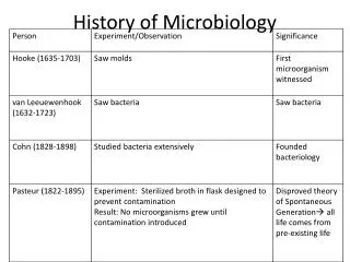
Escherichia coli, originally known as Bacterium coli commune, was identified in 1885 by Theodor Escherich. E. coli is widely distributed in the intestine of humans and warm-blooded animals and is the predominant facultative anaerobe in the bowel and part of the essential intestinal flora that maintains the physiology of the healthy host . • E. coli is a member of the family Enterobacteriaceae, which includes many genera, including known pathogens such as Salmonella, Shigella, and Yersinia. • Most strains of E. coli are not regarded as pathogens, but they can be opportunistic pathogens that cause infections in immunocompromised hosts. • If fecal coliform counts over 200 colonies/100 ml of water sample in the river, there is a greater chance that pathogenic organisms are also present. Such waters has a greater chance to cause diseases by infecting through cuts in the skin, the nose, mouth, or the ears. Diseases and illness such as typhoid fever, hepatitis, gastroenteritis, dysentery, and ear infections can be borned by waters with high fecal coliform counts. There are also pathogenic strains of E. coli that when ingested, causes gastrointestinal illness in healthy humans
In 1892, Shardinger proposed the use of E. coli as an indicator of fecal contamination. This was based on the premise that the pathogens are relatively scarce in water, making them difficult and time-consuming to monitor directly but the E. coli is abundant in human and animal feces and not usually found in other niches. And also. There are correlation between fecal coliform counts and the probability of contracting a disease from the water. Furthermore, since E. coli could be easily detected by its ability to ferment glucose (later changed to lactose), it was easier to isolate than known gastrointestinal pathogens. Hence, the presence of E. coli in food or water became accepted as indicative of recent fecal contamination and the possible presence of frank pathogens. Although the concept of using E. coli as an indirect indicator of health risk was sound, it was complicated in practice, due to the presence of other enteric bacteria like Citrobacter, Klebsiella and Enterobacter that can also ferment lactose and are similar to E. coli in phenotypic characteristics, so that they are not easily distinguished. As a result, the term "coliform" was coined to describe this group of enteric bacteria.
Organisme Indikator Syarat organisme indikator : 1. Keberadaannya dapat terdeteksi pada semua sampel yang akan diperiksa, 2. Pertumbuhan dan jumlahnya memiliki korelasi negatif langsung dengan kualitas sampel, 3. Dapat dideteksi dan dihitung jumlahnya dengan mudah dan dalam waktu singkat, serta dapat dibedakan dengan jelas dari organisme lain, 4. Memiliki resistensi yang tinggi terhadap lingkungan luar (di luar habitat aslinya) agar dapat diisolasi. (Jay 2001: 387 & 389)
Coliform is not a taxonomic classification but rather a working definition used to describe a group of Gram-negative, facultative anaerobic rod-shaped bacteria that ferments lactose to produce acid and gas within 48 h at 35°C. • In 1914, the U.S. Public Health Service adopted the enumeration of coliforms as a more convenient standard of sanitary significance. • Although coliforms were easy to detect, their association with fecal contamination was questionable because some coliforms are found naturally in environmental samples. This led to the introduction of the fecal coliforms as an indicator of contamination. • Fecal coliform, first defined based on the works of Eijkman is a subset of total coliforms that grows and ferments lactose at elevated incubation temperature, hence also referred to as thermotolerant coliforms. Fecal coliform analyses are done at 45.5°C for food testing, except for water, shellfish and shellfish harvest water analyses, which use 44.5°C
The fecal coliform group consists mostly of E. coli but some other enterics such as Klebsiella can also ferment lactose at these temperatures and therefore, be considered as fecal coliforms. The inclusion of Klebsiella spp. in the working definition of fecal coliforms diminished the correlation of this group with fecal contamination. As a result, E. coli has reemerged as an indicator, partly facilitated by the introduction of newer methods that can rapidly identify E. coli. Currently, all 3 groups are used as indicators but in different applications. Detection of coliforms is used as an indicator of sanitary quality of water or as a general indicator of sanitary condition in the food-processing environment. Fecal coliforms remain the standard indicator of choice for shellfish and shellfish harvest waters; and E. coli is used to indicate recent fecal contamination or unsanitary processing.
Almost all the methods used to detect E. coli, total coliforms or fecal coliforms are enumeration methods that are based on lactose fermentation. • The most probable number test, also called the multiple tube fermentation assay, is a statistical, multi-step assay consisting of presumptive, confirmed and completed phases. It is an alternative to plate counts or membrane techniques, especially for samples with higher turbidity. The MPN procedure is a tube-dilution method using a nutrient-rich medium, which is less sensitive to toxicity and supports the growth of environmentally-stressed organisms. The MPN method detects and estimates the bacteria in water samples (and can be applied to foods and soils) by the multiple fermentation tube technique. The number of bacteria per 100 ml of sample is estimated by the use of probability tables. • In the assay, serial dilutions of a sample are inoculated into broth media. Analysts score the number of gas positive (fermentation of lactose) tubes, from which the other 2 phases of the assay are performed and then uses the combinations of positive results to consult a statistical tables, to estimate the number of organisms present. • Typically only the first 2 phases are performed in coliform and fecal coliform analysis, while all 3 phases are done for E. coli.
The 3-tube MPN test is used for testing most foods. The 5-tube MPN is used for water, shellfish and shellfish harvest water testing and there is also a 10-tube MPN method that is used to test bottled water or samples that are not expected to be highly contaminated. • There is also a solid medium plating method for coliforms that uses Violet Red Bile Agar, which contains neutral red pH indicator, so that lactose fermentation results in formation of pink colonies. There are also membrane filtration tests for coliform and fecal coliform that measure aldehyde formation due to fermentation of lactose. This chapter also includes variations of above tests that use fluorogenic substrates to detect E. coli, special tests for shellfish analysis, a brief consideration of bottled water testing and a method for testing large volumes of citrus juices for presence of E. coli in conjunction with the Juice HACCP rule.
Sampling Procedures It is best to use glass bottles but certain types of plastic bottle can be used. Add 4-5 drops of sodium thiosulfate solution (100gr/lt) to each clean sample bottle if the sample contains any residual chlorine. Remove the stopper or cap just before sampling and avoid touching the inside of the cap. If sampling by hand, use gloves and hold the bottle near its base. Plunge it (opening downward) below the water surface, then turn the bottle underwater into the current and away from you. Avoid sampling the water surface because the surface film often contains greater numbers of fecal coliform bacteria than is representative of the river. Also, avoid sampling the sediments for the same reason, unless this is intended. The same general sampling procedures apply when using the extended rod sampler. When collecting samples, leave some space in the sample container (an inch or so) to allow mixing of the sample before-pipetting. It is a good idea to collect several samples room any single location on the river to minimize the variability that comes with sampling for bacteria. Ideally, all samples should be tested within one hour of collection. If this is not possible, the sample bottles should be placed in ice and tested within six hour
Uji Koliform dengan medium standar MTF (Gaudy & Gaudy 1981) Sampel I. UJI PENDUGA Asam + Gas 5 ml 0.5 ml 0.05 ml Inkubasi 24--48 jam 35° C Positif koliform LBG LBT LBT 5ml 5ml 5ml
II. Uji Penguat 1 ose Inkubasi 24--48 jam 35° C Gas LB positif BGLB Positif koliform
III. Uji Pelengkap Koloni hijau metalik: positif E.coli koloni merah: positif koliform pengecatan Gram Streak 24 jam Streak Inkubasi 24 jam 35° C NA EA BGLB positif 1 ose Asam + gas 24--48 jam LB
The presumptive tests are designed to grow the target bacteria. The media used in the confirmed tests are designed to validate the growth of target bacteria in the presumptive test. • Confirmed test conditions are usually more stringent than presumptive conditions. Thus, the presumptive test provides a preliminary estimate of bacterial density based on enrichment in minimally restrictive tube media. The results of this test are never used without further analysis; the MPN must be carried through the Confirmed Test for valid results. • The MPN per 100 ml is calculated from the MPN table based upon the Confirmed Test results. This value is based on the number of positive and negative results observed when five 10 ml, five 1.0 ml and five 0.1 ml volumes of sample are tested in confirmed/completed tests. For example, if 3 of 5 tubes in the first series (10 ml) were positive, 2 of 5 were positive in the second series (1.0 ml) and 1 of 5 positive in the third series (0.1 ml) for confirmed tests, then the pattern is read as 3-2-1. Referring to the MPN index, 3-2-1 implies that the most probable number of bacteria is 17/100 ml. However, the actual range may be between 7 and 40 (95% confidence).
Substrat kromogenik-fluorogenik(Manafi 1991; Manafi 1996; Jay 2001) • Contoh substrat kromogenik untuk deteksi koliform: • o-nitrophenyl-β-D-galactopyranoside (ONPG) dan 5-bromo-4-chloro-3-indolyl--D-galactopyranoside (XGAL). • Contoh substrat fluorogenik untuk deteksi Escherichia coli: • 4-methylumbelliferyl--D-glucoronide (MUG). + sample Medium ± 24 jam • Kromogenik Perubahan warna medium • Fluorogenik Perpendaran medium • (bila dipapar UV) Hidrolisis substrat oleh enzim spesifik
Fluorocult LMX broth Mengandung: Triptosa, triptofan, natrium klorida, sorbitol, K2HPO4, KH2PO4, garam sodium lauryl sulfat , 5-bromo-4-chloro-3-indolyl--D-galactopyranoside (XGAL), 4-methylumbelliferyl --D-glucoronide (MUG), dan 1-isopropyl--D-1-thiogalactopyranoside (IPTG). IPTG (Inducer) XGAL (Substrat kromogenik) -galaktosidase Aglycone bebas Koliform Cat indigo Warna biru kehijauan
MUG Substrat fluorogenik Triptofan asam amino -glukuronidase 4-MU Escherichia coli Sinar UV berpendar triptofanase Indol Escherichia coli Reagen kovac cincin merah
Uji Koliform pada Fluorocult LMX broth Warna biru kehijauan 5 ml 0.5 ml 0.05 ml Inkubasi 24--48 jam 35° C Fluorocult LMX broth tunggal Fluorocult LMX broth tunggal Fluorocult LMX broth ganda Positif koliform
UV light Uji indole berpendar E.coli Fluorocult LMX Positif koliform Terbentuk cincin merah
Pengecatan Gram Cuci Fiksasi Cuci NA dari fluorocult LMX broth Akuades steril Cuci Fiksasi Amati Cuci NA dari Endo agar Akuades steril
Uji Indol Inkubasi 24--48 jam 35° C NA dari Endo agar Tripton 1% Terbentuk cincin merah Positif E. coli.