Cell Structure and Function
Cell Structure and Function. Chapter 5. Cell Theory. Every organism is composed of one or more cells Cell is smallest unit having properties of life Life arises from growth and division of single cells (cells come from pre-existing cells). Cell. Smallest unit of life

Cell Structure and Function
E N D
Presentation Transcript
Cell Structure and Function Chapter 5
Cell Theory • Every organism is composed of one or more cells • Cell is smallest unit having properties of life • Life arises from growth and division of single cells (cells come from pre-existing cells)
Cell • Smallest unit of life • Can survive on its own or has potential to do so • Is highly organized for metabolism • Senses and responds to environment • Has potential to reproduce
Cell size • Most cells range in diameter from ~1 µm-100 µm • (1 µm = 10-6 m) • Why are cells so small?
Why Are Cells So Small? • Surface-to-volume ratio • The bigger a cell is, the less surface area there is per unit volume • Above a certain size, material cannot be moved in or out of cell fast enough
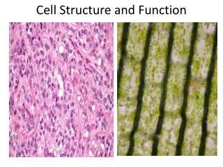
Structure of Cells Two Main Classes of Cells • Prokaryotic: simple organization with no nucleus or organelles. • Eukaryotic: highly developed membrane system, DNA encapsulated in nucleus, various organelles.
Prokaryotic Cells • Archaea and Bacteria • Contain no nucleus or organelles • Made up of: • Cell membrane • Cell wall • Cytoplasm • Nucleoid region (DNA) • Ribosomes • May contain • Pili • Capsule • flagella
Prokaryotic Structure pilus cytoplasm with ribosomes DNA flagellum capsule cell wall plasma membrane
Eukaryotic cells: Plant Cell Animal Cell
Eukaryotic Cellular Organization • Plasma membrane: • Encapsulates the cell, lipid bilayer • Nucleus: • Contains the genetic material • Cytoplasm: • Everything in between • Contains various organelles
Organelles • Small specialized membrane-bound compartments found in EUKARYOTIC cells • Common organelles • Nucleus • Endoplasmic reticulum (ER) • Golgi body • Lysosomes • Vesicles • Vacuoles • Mitochondria • Chloroplasts
Non-Membrane Structures • Cytoskeleton • Ribosomes
Membranes • Composed of phospholipid bilayer • Double layer of phospholipids • Organized with their hydrophobic tails pointing inward. • The hydrophilic heads point out in the solvent • Also contains proteins • Channels allow molecules in or out of the cell either by passive flow or active pumping. • Receptors bind signaling molecules given off the other cells and trigger changes within the cell.
lipid bilayer fluid fluid one layer of lipids one layer of lipids Membranes
Functions of Nucleus • Keeps the DNA molecules of eukaryotic cells separated from metabolic machinery of cytoplasm • Makes it easier to organize DNA and to copy it before parent cells divide into daughter cells
Components of Nucleus Nuclear envelope • Chromatin • DNA + Proteins • Nucleolus • Site of ribosome production Figure 4.11bPage 62
Nuclear Envelope • Double lipid bilayer • Contains Nuclear pores – involved in transporting material in and out of nucleus Nuclear pore bilayer facing cytoplasm Nuclear envelope bilayer facing nucleoplasm Figure 4.12bPage 63
Endomembrane System • Group of related organelles in which lipids are assembled and new polypeptide chains are modified • Products are sorted and shipped to various destinations • A system for the transport & processing of complex molecules running from the nucleus to the cell’s surface
Components of Endomembrane System • Nuclear Envelope • Smooth & Rough Endoplasmic Reticulum (ER) • Golgi Body • Vesicles • Cell membrane
Endoplasmic Reticulum • In animal cells, continuous with nuclear membrane • Extends throughout cytoplasm • Two regions • Rough (RER)– Produces and processes proteins • Smooth (SER) – Lipid assembly
Golgi Body • Puts finishing touches on proteins and lipids that arrive from ER • Packages finished material for shipment to final destinations • Material arrives and leaves in vesicles budding vesicle
Vesicles • Membranous sacs that move through cytoplasm • Lysosomes - Contain hydrolytic enzymes to break down wastes (garbage & recycling); require low pH (~5-6) • Peroxisomes - contain enzymes to break down dangerous oxygen molecules • Transport vesicles - Carry proteins, etc. to different parts of cell • Vacuoles - storage (water, nutrients, wastes); very large vacuole in plants
Mitochondria • ATP-producing powerhouses - Site of cellular respiration • Membranes form two distinct compartments • Matrix: Inner compartment • Cristae: folds of the inner membrane
Specialized Plant Structures • Cell Wall • Chloroplast • Central Vacuole
Chloroplasts • Found only in plants and are the location of photosynthesis • Convert sunlight into ATP which is then used to convert water and CO2 into sugar. • Pigments contained within trap light and harness its energy
Cell Wall • Made of cellulose (polysaccharide made of glucose subunits) • Makes plant cells rigid • Stores water, nutrients, wastes • Maintains pressure and rigidity of cell Central Vacuole
Mitochondrial and Chloroplast Origins • Mitochondria resemble bacteria • Have own DNA, ribosomes • Divide on their own • Endosymbiosis: May have evolved from ancient bacteria that were engulfed but not digested
Mitochondrial and Chloroplast Origins: Endosymbiosis http://evolution.berkeley.edu/evolibrary/images/endosymbiosis/endosymbiosis.gif
Mitochondrial and Chloroplast Origins: Endosymbiosis http://faculty.ircc.edu/faculty/tfischer/images/endosymbiosis.jpg
Cytoskeleton • Present in all eukaryotic cells • Basis for cell shape and internal organization • Allows organelle movement within cells and, in some cases, cell motility
a. Microtubule Microtubules Tubulin subunits • Composed of tubulin • Arise from Centrosomes • Centrioles in animal cells • Involved in shape, motility, cell division 25 nm
c. Microfilament Microfilaments • Thinnest elements • Composed of actin • Take part in movement, formation, and maintenance of cell shape 5–7 nm Actin subunit
b. Intermediate filament Intermediate Filaments • Only in animal cells of certain tissues • Most stable cytoskeletal elements • Six known groups Each green line is an intermediate filament protein 8–12 nm Figure 4.21Page 71
Motor Proteins • Kinesins and dyneins move along microtubules • Myosins move along microfilaments kinesin microtubule Figure 4.24b, Page 72
Flagella and Cilia • Both are used for cellular propulsion • Cilia are also used for food collection or to move substances across a cell’s surface • Cilia are short and more numerous, while flagella are longer (>2mm), only 1 or 2 are found per cell
Flagella and Cilia • Both are constructed similarly w/ a central microtubule bundle containing a “9+2” arrangement: Cross section of flagellum Micrograph of flagellum Plasma membrane Dynein arm Two central microtubules Central sheath Spoke Links of the connective system
Cilia and Flagella Flagella beat in smooth, S-shaped waves that travel from base to tip. Tip Base Cilia beat in an oarlike power stroke (dark orange) followed by a recovery stroke (light orange).
Ribosomes • Complex made up of proteins and RNA • Site of protein synthesis • Made up of two subunits, produced in the nucleolus • Subunits are then exported into cytoplasm • In the cytoplasm they are assembled into a functional ribosome
Cell-to-Cell Junctions • Plants • Plasmodesmata • Animals • Tight junctions • Anchoring junctions • Gap junctions plasmodesma
Functions of Cellular Junction • Anchoring junctions “weld” cells together • Desmosomes and adherens • Tight junctions – water tight seal between cells • Seal spaces and fuse membranes • Gap junctions – “channels” between cells • allow for exchange/communication between one cell and the next
Cells Channel in a complex of proteins Intermediate filaments Plaque Gap junction: Cylindrical arrays of proteins form direct channels that allow small molecules and ions to flow between the cytoplasm of adjacent cells. Anchoring junction: Adjoining cells adhere at a mass of proteins (a plaque) anchored beneath their plasma membrane by many intermediate filaments (adherens junction) or microfilaments (desmosome) of the cytoskeleton. Tight junction: Tight connections form between adjacent cells by fusion of plasma membrane proteins on their outer surfaces. A complex network of junction proteins makes a seal tight enough to prevent leaks of ions or molecules between cells. Fig. 5-26, p. 114
Extracellular Matrix • Collagen proteins • Tensile strength and elasticity • Proteoglycans • Interlinkage • Changes consistency (jellylike to hard and elastic) • Fibronectins • Connect cells via integrins